 January 2017
On the Case
By Pramod Gupta, MD, and Francisco Garcia-Morales, MD
Radiology Today
Vol. 18 No. 1 P. 32
History
A 67-year-old man was admitted to the hospital for atypical chest pain and shortness of breath. He had a history of hypertension, COPD with a long smoking history, and alcohol dependence. He underwent cardiac catheterization to assess for coronary artery disease. The left coronary artery could not be engaged despite multiple attempts. A CT angiogram was performed next to rule out anomalous origin of left coronary artery.
Findings
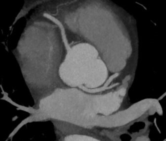
Axial CT maximum intensity projection (MIP) image shows anomalous origin of the left main coronary artery from the noncoronary sinus (Figure 1), which runs leftward posterior to the aorta with further normal expected course.
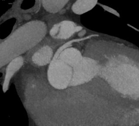
Coronal oblique MIP image (Figure 2) shows the normal diameter of the ostium of the left main coronary artery without an acute angulation.
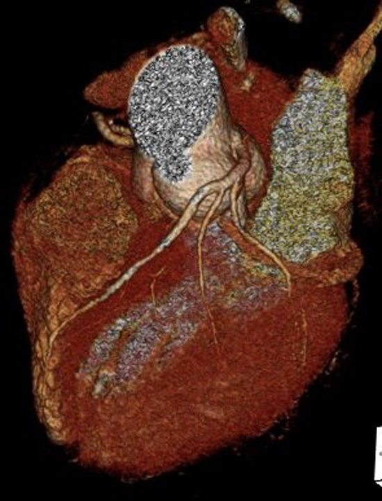
3D volume rendered image (Figure 3) shows the posterior course of the left main coronary artery and then normal branching.
Diagnosis
Anomalous origin of the left main coronary artery from the posterior noncoronary sinus of Valsalva with benign course.
Discussion
Anomalous origin of the coronary arteries in those without congenital heart disease is relatively rare and occurs in less than 1% of all adult patients undergoing coronary angiography. While assessing the anomalous coronary artery, it is very important to determine whether it has a benign or malignant (hemodynamically significant) course. Because of abnormal anatomic takeoff, the anomalous coronary artery may have an intramural course and be associated with a slitlike ostium at origin. These patients are at an increased risk of acute myocardial infarction and sudden death due to deficiencies in coronary flow secondary to either impaired flow through the slitlike ostium or compression between the aorta and pulmonary trunk (interarterial course). For these hemodynamically significant anomalies, open surgical intervention is usually required. Cardiac CT is an excellent modality in depicting the morphology of the coronary artery lumen three-dimensionally.
This case, which shows anomalous origin of the left main coronary artery from the noncoronary sinus of Valsalva, is extremely rare and has been previously reported only in a few case reports. Most of the cases were diagnosed incidentally in asymptomatic patients. Due to the extreme rarity of this anomaly, the natural history and risk of adverse events related to it are unknown.
In this case, the anomalous artery had normal ostial diameter and a benign retroaortic course.
— Pramod Gupta, MD, and Francisco Garcia-Morales, MD, are staff radiologists at the Dallas VA Medical Center.
Resources
1. Liberman L, Pass RH, Kaufman S, Hordof AJ, Printz BF, Prakash A. Left coronary artery arising from the non-coronary sinus: a rare congenital coronary anomaly. Pediatr Cardiol. 2005;26(5):672-674.
2. Shriki JE, Shinbane JS, Rashid MA, et al. Identifying, characterizing, and classifying congenital anomalies of the coronary arteries. Radiographics. 2012;32(2):453-468.
3. Garg A, Ogilvie BC, McLeod AA. Images in Cardiology. Anomalous origin of the left coronary artery from the non-coronary sinus of Valsalva. Heart. 2000;84(2):136.
4. Nowak B, Kupferwasser I, Mayer E, et al. Images in cardiovascular medicine. Anomalous origins of the left main coronary artery from the noncoronary sinus and of the right coronary artery from the left sinus of Valsalva. Circulation. 1997;96(8):2731-2732.
