By Rahul V. Pawar, MD
History
A 44-year-old man scheduled for brain MRI has a long history of seizures (simple, partial), including convulsions. MRI was performed as part of a comprehensive semiologic workup.
Finding
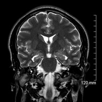
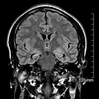
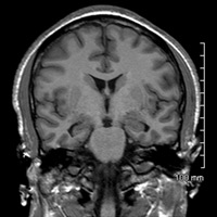
Coronal FLAIR and T2- and T1-weighted imaging demonstrate volume loss of the right hippocampal formation. FLAIR imaging confirms mild hyperintensity within the hippocampal gyrus; secondary enlargement of the parahippocampal fissure is dually noted.
Diagnosis
Right-sided mesial temporal sclerosis (MTS)
Discussion
Neuroimaging plays a critical role in evaluating patients with refractory epilepsy. Occasionally, anatomic abnormalities are detected that may be amenable to surgical resection (potentially curative).
MTS is a well-known cause of intractable temporal lobe epilepsy. Histologically, MTS is characterized by neuronal loss/gliosis of the hippocampus. In moderate and severe cases, volume loss of the ipsilateral fornix and mammillary body can be seen. As part of the Papez circuit, the fornix is critical regarding the formation of memory, with damage or disease resulting in anterograde amnesia.
A complete semiologic evaluation (seizure workup) rests on clinical, electroencephalograph, and neuroimaging data. The power of neuroimaging cannot be overstated. A powerful, efficient MRI is critical to ensuring that clear, sharp images are generated. Identifying MTS can be challenging since the anatomy involved is delicate and (positive) imaging findings are often subtle. This patient was scanned on a Philips Achieva 1.5T magnet that utilizes FreeWave 16/32-channel architecture that supports a wide range of multichannel coils designed to achieve fast imaging and high-spatial resolution. Anxious and motion-prone patients can benefit from rapid acquisition.
MRI has a significant diagnostic and prognostic impact on patients with epilepsy. Structural abnormalities aside from MTS, such as malformations of cortical development, can be associated with medically refractory seizures. In addition to high-resolution imaging, advances and collaboration between allied fields in neuroscience have led to the development of quantitative methods or statistical parametric mapping of brain anatomy.
Recently published data regarding MTS and preoperative neuroimaging have shown promise; diffusion tensor imaging may be able to detect putative abnormalities in connectivity within the medial temporal lobes, although much work remains in this field.
— Rahul V. Pawar, MD, is an attending neuroradiologist at Saint Barnabas Medical Center in Livingston, New Jersey, and a consultant for Philips Healthcare North America.
 |
|
 |
|
 |
- Rastogi S, Lee C, Salamon N. Neuroimaging in pediatric epilepsy: a multimodality approach. Radiographics. 2008;28:1079-1095.
- Thomas AG, Koumellis P, Dineen RA. The fornix in health and disease: an imaging review. Radiographics. 2011;31(4):1107-1121.
- Dupont S, Baulac M. [Contribution of MRI to the exploration of partial refractory epilepsy.] Rev Neurol (Paris). 2004;160 Spec No 1:5S91-5S97.
- Zeineh MM, Holdsworth S, Skare S, Atlas SW, Bammer R. Ultra-high resolution diffusion tensor imaging of the microscopic pathways of the medial temporal lobe. Neuroimage. 2012;62(3):2065-2082.
Submission Instructions
Submit cases directly to Rahul V. Pawar, MD, DABR (section editor for “On the Case”) at rvp325@yahoo.com. Cases submitted should be relevant and interesting. All modalities and subspecialties within radiology are equally considered.
Case submission entails two PowerPoint slides:
SLIDE 1
a. History (one-line phrase)
b. Two to five high-quality images in JPEG format without annotations
c. Name(s) of the author(s) (three maximum) and respective institutions
SLIDE 2
a. Diagnosis
b. Concise bulleted discussion (one to two lines each), including the following: pertinent clinical history, diagnostic imaging findings, differential diagnoses, treatment (if applicable)
c. Two to three relevant and current references, preferably citing peer-reviewed radiology literature
Department of Radiology, Division of Neuroradiology
Saint Barnabas Medical Center/Barnabas Ambulatory Care Center