“Chasing the Dragon’
History and Course of Illness
A 43-year-old Chinese man with an unknown past medical history was brought to the emergency department with altered mental status. Although vital signs were stable, his neurological function deteriorated rapidly; ultimately, he became nonverbal, unable to perform his activities of daily living and follow commands. A physical exam revealed spastic upper extremities and minimal lower extremity withdrawal to pain. Basic metabolic panel, complete blood count, and cerebrospinal fluid analysis were all negative. Urine toxicology screen was positive for methadone and opiates. Both CT and MR imaging of the head were performed. Coenzyme Q, vitamin E, and a GABA-receptor agonist were initiated. A percutaneous gastrostomy tube was placed, and the patient was transferred to a chronic care facility for neurorehabilitation.
Findings
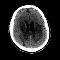
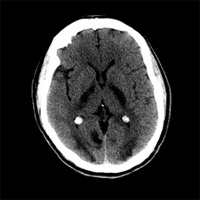
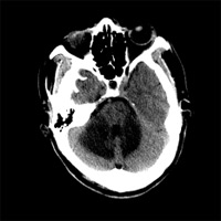
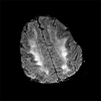
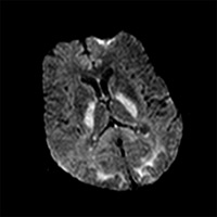
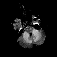
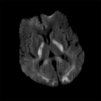
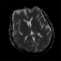
Noncontrast CT scan of the supra-tentorial brain reveals symmetric hypoattenuation within the centrum semiovale (Figure 1A), posterior limbs of the internal capsules, and parietooccipital white matter (Figure 1B). Within the posterior fossa, there is symmetric involvement of the cerebellar white matter, particularly within the middle cerebellar peduncles (Figure 1C). Axial fluid-attenuated inversion recovery (FLAIR) sequences from MR imaging demonstrates an equivalent distribution of hyperintense signal (Figures 2A and 2B), with additional signal abnormality involving the corticospinal tracts and middle cerebellar peduncles (Figure 2C). Diffusion-weighted imaging (DWI) reveals the presence of abnormal restricted diffusion within the posterior limbs of the internal capsules and posterior white matter (Figures 3A and 3B).
Diagnosis
Toxic leukoencephalopathy secondary to heroin vapor inhalation (aka, “chasing the dragonâ€)
Discussion
To avoid the hazards of intravenous drug use, an alternative and potentially lethal method of abusing heroin was conceived in China during the early 20th century. Inhaling a vaporized form of heroin (or any morphine derivative) supposedly provides a unique “high†without using hypodermic needles. Heating the diacetylmorphine hydrochloride salt or powder form of heroin on aluminum foil liquefies it into a reddish-brown gelatinous substance. The exothermic reaction yields a vapor suitable for inhalation through various contraptions.1
Profound damage occurs to the central nervous system in the form of toxic spongiform leukoencephalopathy and vacuole formation. CT demonstrates symmetric hypoattenuation within the centrum semiovale, posterior limbs of the internal capsules, parieto-occipital, and cerebellar white matter. Within the midbrain, there is involvement of the medial lemnisci and corticospinal tracts. Areas that are notably spared include the subcortical U-fibers, anterior limbs of the internal capsules, and dentate nuclei. MR imaging corroborates what can be appreciated on CT, demonstrating symmetric high signal within the affected white matter on T2 and FLAIR sequences. DWI may also be positive, demonstrating hyperintense signal on the trace images (Figure 3A), with corresponding deficit on the apparent diffusion coefficient map (Figure 3B). Although the findings on DWI are suggestive of typical hypoxic-ischemic insult, MR spectroscopy data have confirmed the presence of lactate confined to the affected white matter, raising the possibility of mitochondrial dysfunction. Furthermore, in a subset of patients treated with antioxidants and coenzyme Q, there has been documented recovery of substantial neurological function. Biopsy specimens have also revealed a significant degree of axonal sparing and a lack of Wallerian degeneration, which may also serve to explain why some level of neurological dysfunction is reversible.2, 3
Clinically, the spectrum of findings may be broad. Patients may present with altered mental status and pseudobulbar signs such as slurred speech or swallowing/chewing difficulties. Toward the more catastrophic end, some may develop aphasia, spastic quadraparesis, and progress to coma and death.
An awareness of this disease process is essential. Though “chasing the dragon†can be traced back to 1920s China, the phrase carries more than just historical relevance. This potentially devastating method of opioid abuse has established a foothold across a wide demographic, including homeless and drug-addicted youths in the United States.┬ Aside from a thorough history and neurological exam, neuroimaging can be instrumental in suggesting the diagnosis. MRI delineates the distribution of white matter involvement particularly well. At the very least, supportive care and vitamin supplementation may help mitigate the effects on the patient’s nervous system.
— Rahul V. Pawar, MD, is a board-certified diagnostic radiologist completing a fellowship in neuroradiology at New York University Langone Medical Center.
References
 |
 |
| Figure 1A — Noncontrast CT scan demonstrates symmetric hypoattenuation within the periventricular white matter and centrum semiovale. | Figure 1B — Noncontrast CT scan┬ demonstrates symmetric hypoattenuation within the posterior limbs of the internal capsules and parietooccipital white matter. |
 |
 |
| Figure 1C — Noncontrast CT scan through the posterior fossa demonstrates symmetric hypoattenuation within the middle cerebellar peduncles. | Figure 2A — In Figures 2A and 2B, axial fluid-attenuated inversion recovery (FLAIR) sequences demonstrates an equivalent distribution of hyperintense signal (centrum semiovale, posterior limbs of the internal capsules, and parieto-occipital white matter) |
 |
 |
| Figure 2B | Figure 2C — FLAIR sequence demonstrates signal hyperintensity involving the corticospinal tracts and middle cerebellar peduncles. |
 |
 |
| Figure 3A — Trace diffusion image demonstrates hyperintense signal within the posterior limbs of the internal capsules and parietooccipital white matter. | Figure 3B — ADC map confirms the presence of abnormal restricted diffusion with the corresponding hypointense signal. |
REFERENCES
- Gossop M, Griffiths P, Strang J. Chasing the dragon: Characteristics of heroin chasers. Br J Addict. 1988;83(10):1159-1162.
- Keogh CF, Andrews GT, Spacey SD, et al. Neuroimaging features of heroin inhalation toxicity: “Chasing the dragon.†AJR Am J Roentgenol. 2003;180(3):847-850.
- Kriegstein AR Shungu DC, Millar WS, et al. Leukoencephalopathy and raised brain lactate from heroin vapor inhalation (“chasing the dragonâ€). Neurology. 1999;53(8):1765-1773.
ON THE CASE submission requirements
- Cases should have clinical relevance and clear radiological findings.
- Sections should include a title, history and course of illness, findings, diagnosis, and discussion.
- Maximum word limit should not exceed 800. At least three references are recommended.
- Cases may be submitted from any radiological subspecialty and imaging modality.
- Figures must be high-quality JPEG or TIFF images and labeled for ease of reference. Please keep images in their native format, without the addition of arrows or other means of highlighting the key findings.