By Rahul V. Pawar, MD, DABR; David Dunaway, DO;
and Lyle R. Gesner, MD
History
Seizures (successfully treated with antiepileptic medications)
Description
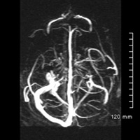
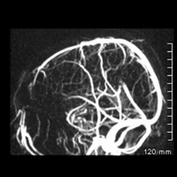
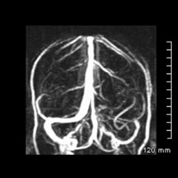
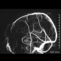
Persistent drainage pathways that connect the confluence of sinuses with the sigmoid sinus/internal jugular vein bilaterally. Atretic left transverse/sigmoid sinus segments (incidental).
Diagnosis
Persistent occipital sinuses
Discussion
• Aside from understanding the various limitations/pitfalls of MR venography, recognizing anatomic variants of major venous drainage is critical and surgically relevant.
• “Flow gaps” can be seen in up to 30% of normal MR scans typically within the hypoplastic transverse sinus, principally as a function of slow intravascular flow, in-plane flow, and/or complex flow patterns. Hypoplasia of the transverse or sigmoid sinus may be associated with alternative drainage pathways (eg, persistent occipital sinus).
• Occipital sinuses usually communicate cranially at the torcular Herophili and caudally at the foramen magnum, where a multitude of variations may exist.
• Occipital sinuses have been reported in up to 10% of normal subjects and may be associated with atretic transverse/sigmoid sinus segments and internal jugular vein. Correlation with CT may bolster confident diagnosis by demonstrating a smaller caliber sigmoid sinus groove and/or jugular foramen.
• Occipital venous network is believed to regress once most of the venous flow passes through the major dural sinuses when children assume the upright position.
• Relevance: (Accidental) discovery of a persistent occipital sinus during posterior fossa surgery can result in catastrophic hemorrhage and/or occlusion of the occipital sinus that may lead to venous thrombosis/infarction.
— Rahul V. Pawar, MD, DABR, is a radiologist at Saint Barnabas Medical Center in Livingston, New Jersey.
— David Dunaway, DO, is a radiology resident at Saint Barnabas.
— Lyle R. Gesner, MD, is the neuroradiology section head at Saint Barnabas.
 |
 |
 |
 |
REFERENCES
- Provenzale JM, Kranz PG. Dural sinus thrombosis: sources of error in image interpretation. AJR Am J Roentgenol. 2011;196(1):23-31.
- Kobayashi K, Suzuki M, Ueda F, Matsui O. Anatomical study of the occipital sinus using contrast-enhanced magnetic resonance venography. Neuroradiology. 2006;48(6):373-379.
- Ayanzen RH, Bird CR, Keller PJ, McCully FJ, Theobald MR, Heiserman JE. Cerebral MR venography: normal anatomy and potential diagnostic pitfalls. AJNR Am J Neuroradiol. 2000;21(1):74-78.
Submission Instructions
Submit cases directly to Rahul V. Pawar, MD, DABR (section editor for “On the Case”) at rvp325@yahoo.com. Cases submitted should be relevant and interesting. All modalities and subspecialties within radiology are equally considered.
Case submission entails two PowerPoint slides:
SLIDE 1
a. History (one-line phrase)
b. Two to five high-quality images in JPEG format without annotations
c. Name(s) of the author(s) (three maximum) and respective institutions
SLIDE 2
a. Diagnosis
b. Concise bulleted discussion (one to two lines each), including the following: pertinent clinical history, diagnostic imaging findings, differential diagnoses, treatment (if applicable)
c. Two to three relevant and current references, preferably citing peer-reviewed radiology literature
Department of Radiology, Division of Neuroradiology
Saint Barnabas Medical Center/Barnabas Ambulatory Care Center