By Rayhan S. Hai, MD; Pramod Gupta, MD;
and Kanwar Singh, MD
History
A 36-year-old woman with no past medical history presented with the complaint of central chest pain over the past six months. The initial chest X-rays were normal, though the patient continued to complain of worsening pain and a sensation of chest swelling.
A CT scan of the chest six months after initial symptoms revealed a sternal mass. Biopsy findings were consistent with plasmacytoma.
Findings
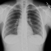
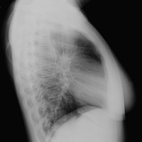
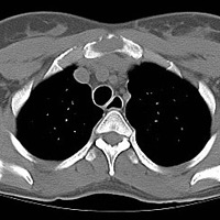
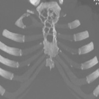
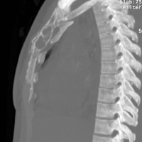
The initial chest X-rays (Figures A and B) are normal. The axial CT image (Figure C) through the chest demonstrates an expansile soft tissue density lytic lesion in the sternum. The mass is surrounded posteriorly by a thin rim of cortical bone, though it appears to disrupt the cortex anteriorly. The coronal (Figure D) and sagittal (Figure E) maximum-intensity projection CT scan images demonstrate the multilobulated “bubbly” appearance of the lesion, a classic feature.1
Differential Diagnosis
Chondrosarcomas are the most common primary tumor of the sternum. They generally are painless and demonstrate chondroid matrix mineralization.1 Lymphomatous masses can appear similar to a solitary bone plasmacytoma (SBP), though the cortex is generally intact around the bone.1 Solitary expansile thyroid or renal cell carcinoma metastases may mimic the appearance of an SBP.1
Discussion
SBP is a rare, localized proliferation of plasma cells centered in the bone marrow. The diagnosis is made when biopsy reveals a monoclonal population of plasma cells and systemic myeloma is excluded.2
Patients often present clinically with pain at the site of the lesion, as in this case. The most common site for occurrence is the axial skeleton, especially the vertebrae.3
About 30% of patients with SBP will have multiple myeloma at presentation. Of those who don’t have multiple myeloma, the treatment of choice is localized radiation.1 However, the majority of patients with SBP eventually will develop multiple myeloma.3
— Rayhan S. Hai, MD, is an internist at The University of Texas Southwestern Medical Center in Dallas.
— Kanwar Singh, MD, is an internist at The University of Texas Southwestern Medical Center.
— Pramod Gupta, MD, is a radiologist at the Dallas VA Medical Center.
 |
 |
| Figure A | Figure B |
 |
 |
| Figure C | Figure D |
 |
|
| Figure E |
- Restrepo CS, Martinez S, Lemos DF, et al. Imaging appearances of the sternum and sternoclavicular joints. Radiographics. 2009;29(3):839-859.
- Dimopoulos MA, Kiamounis C, Moulopoulos LA. Solitary plasmacytoma of bone and extramedullary plasmacytoma. Hematol Oncol Clin North Am. 1999;13(6):1249-1257.
- Soutar R, Lucraft H, Jackson G, et al. Guidelines on the diagnosis and management of solitary plasmacytoma of bone and solitary extramedullary plasmacytoma. Br J Haematol. 2004;124(6):717-726.
Submission Instructions
- Cases should have clinical relevance and clear radiological findings.
- Seconds should include a title, history and course of illness, findings, diagnosis, and discussion.
- Word count should not exceed 800. At least three references are recommended.
- Cases may be submitted from any radiological subspecialty and imaging modality.
- Figures must be high-quality JPEG or TIFF images and labeled for ease of reference. Please keep images in their native format, without the addition of arrows or other means of highlighting the key findings.
Submit cases via e-mail to Rahul V. Pawar, MD, at rvp325@gmail.com or to Radiology Today at jknaub@gvpub.com.
Department of Radiology, Division of Neuroradiology
Saint Barnabas Medical Center/Barnabas Ambulatory Care Center