By Sara Ali, MD; Adil Omer, MD; and Dane Blumenthal, MD
History
A 33-year-old Hispanic female with no significant past medical history presented to a breast clinic complaining of a painful left breast mass of one month duration. She reported no family history of breast carcinoma or history of breast surgery. Clinical breast examination revealed a 4 cm mass in the left breast without skin changes. Mammography and breast ultrasound were then performed.
Findings
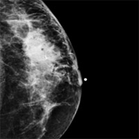
Mammography demonstrated a 4 cm asymmetric density in the upper outer quadrant of the left breast, which persisted on spot compression imaging (Figures A and B).
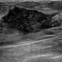
Breast sonography demonstrated a corresponding 4 cm mixed echogenic predominantly hypoechoic mass in the left breast at 1 o’clock position 3 cm from the nipple (Figure C).
Ultrasound guided core needle biopsy of the left breast mass was then performed.
Diagnosis
- Invasive ductal carcinoma
- Invasive lobular carcinoma
- Metastases/lymphoma
- Granulomatous mastitis (tuberculosis/histoplasmosis/sarcoid)
- Posttraumatic hematoma/fat necrosis
- Postsurgical scar
Discussion
Pathology results from left breast mass ultrasound guided core needle biopsy revealed “noncaseating granulomatous inflammation, favor sarcoidosis.”
Sarcoidosis is a multisystemic inflammatory granulomatous disease of unknown etiology. It most commonly affects the lungs and lymphatic system.1 Sarcoid rarely (less than 1%) involves the breast and is particularly unusual in the breast without a history of systemic sarcoid as in the incident case.2 Although breast sarcoid should be considered a diagnosis of exclusion, breast sarcoid should be included as a differential consideration in patients with a breast mass and a history of systemic sarcoid.
It should be kept in mind that the breast imaging manifestations of sarcoid and breast cancer can overlap. Sarcoid involving the breast can present as an irregular spiculated mass on mammography or as a hypoechoic mass on ultrasound.3 Further, MR imaging does not aid in differentiating sarcoid from breast carcinoma as both may present as enhancing masses.
In light of these similar imaging characteristics involving sarcoid and breast carcinoma, image guided core needle biopsy is indicated to differentiate the rare sarcoid from the more prevalent breast carcinoma when evaluating patients with a history of sarcoid and a breast mass found during imaging.
Our report demonstrates a rare case of primary breast sarcoid in a young female with no prior history of systemic sarcoid.
— Sara Ali, MD, is the chief radiology resident at Harlem Hospital Center in New York.
— Adil Omer, MD, is a clinical researcher at Harlem Hospital Center.
— Dane Blumenthal, MD, is an attending radiologist at Harlem Hospital Center.
 |
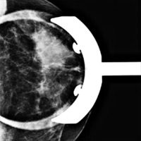 |
| Figure A | Figure B |
 |
|
| Figure C |
References
- American Thoracic Society, the European Respiratory Society, the World Association of Sarcoidosis and Other Granulomatous Disorders. Statement on sarcoidosis. Am J Respir Crit Care Med. 1999;160(2):736-755.
- Isley LM, Cluver AR, Leddy RJ, Baker MK. Primary sarcoid of the breast with incidental malignancy. J Clin Imaging Sci. 2012;2:46.
- Sabaté JM, Clotet M, Gómez A, De Las Heras P, Torrubia S, Salinas T. Radiologic evaluation of uncommon inflammatory and reactive breast disorders. Radiographics. 2005;25(2):411-424.
Submission Instructions
- Cases should have clinical relevance and clear radiological findings.
- Seconds should include a title, history and course of illness, findings, diagnosis, and discussion.
- Word count should not exceed 800. At least three references are recommended.
- Cases may be submitted from any radiological subspecialty and imaging modality.
- Figures must be high-quality JPEG or TIFF images and labeled for ease of reference. Please keep images in their native format, without the addition of arrows or other means of highlighting the key findings.
Submit cases via e-mail to Rahul V. Pawar, MD, at rvp325@gmail.com or to Radiology Today at jknaub@gvpub.com.
Department of Radiology, Division of Neuroradiology
Saint Barnabas Medical Center/Barnabas Ambulatory Care Center