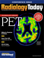
 May 2012
Technology Update: PET
By Dan Harvey
Radiology Today
Vol. 13 No. 5 P. 16
PET is more than a foundation on which to build a hybrid technology. In its basic form, it continues to provide excellent spatial and temporal resolution for clinicians in cardiology, neurology, and oncology. Once deemed expensive, now PET is often considered a cost-effective option in the molecular imaging realm.
But versatility is a main descriptor. PET remains fertile ground for a hybridized seed that can flourish into new modalities. Consider PET/MRI.
Positron
Cardiology Focus
Positron, the Indiana-based, nuclear cardiology-focused molecular imaging company, has developed a cardiac-optimized PET scanner. Attrius, an accurate and cost-effective solution, goes against the market grain, says Chief Technology Officer Joseph Oliverio. “We moved away from PET/CT to create a PET camera optimized for cardiologists that didn’t sacrifice sensitivity for resolution.”
That kind of trade-off existed with early PET scanners for cardiac imaging; Positron went the opposite way, eschewing the trade-off for a system targeted at cardiac imaging. “We perceived a need for a dedicated scanner that imaged the way a cardiologist wants and that provided other substantial benefits,” Oliverio says.
Its product—which went into development in 2006 and was FDA approved in 2009—is designed to provide high-system uptime, reduced radiation exposure, and proprietary cardiac-specific software. In 2010, the device received Frost & Sullivan’s North American Molecular Imaging System New Product Innovation Award.
The Attrius footprint is small enough to fit into a 15- X 20-ft area and costs less compared with PET/CT. Considering the initial price, installation costs, and ongoing maintenance, a PET/CT purchase can be a hard sell for private cardiology practices, Oliverio says. While a PET procedure is more costly than a SPECT exam, a PET deployment in cardiac nuclear medicine can reduce long-term costs, according to the company.
Positron says the system is designed with fewer boards, easier access to detector modules, automated turning features built into the gantry, and coronary artery overlay display. A large list mode buffer enables concurrent acquisition of flow, perfusion, and function in a single scan. The integrated INTEL chipset provides cardiologists quick assessment with reconstruction times of ungated images in less than 7 seconds, Oliverio says.
The system is also designed to use less power. “We designed it for today’s electronics environment. It needs barely any additional air conditioning or power requirements,” Oliverio says. “Also, anything that can be connected by ethernet is accessible through the Internet.”
Further, Attrius enables rapid, dependable studies for larger patients, featuring a 450-lb table capacity. The table can be raised or lowered for easy, comfortable patient loading.
The open architecture fosters new protocol development and customization.
“Neurology is an emerging area, and we see potential to place scanners in neurologists’ offices for brain imaging,” Oliverio says. “But that’s something we might look at down the road.”
GE Healthcare
Suite Solution for Oncology
While Positron focuses on cardiology, GE Healthcare’s new PET technology products focus on oncology. During RSNA 2011, the company introduced Q.Suite, a tool set designed to enhance PET for cancer treatment.
“Assessing activity level, or uptake of cancer cells, that’s the key,” says Vivek Bhatt, GE Healthcare’s general manager for PET. “[The modality’s promise] is repeated imaging and accurately measured activity level. Modalities such as MR and CT visualize the tumor size but don’t reveal the change level.”
The company helps clinicians realize the power of PET. “Measurement of activity level opens it up,” Bhatt says. “Clinicians not only see the tumor and observe metabolic change but are better able to determine if treatment is effective.”
All technology improvements are critical, but one of the most important pieces involves quantitation. “PET imaging’s potential resides in treatment assessment,” Bhatt says. “[The] resulting treatment modification increases the chance of effective therapy.”
Such issues drove development of Q.Suite’s set of hardware and software tools designed to reduce variability and, in turn, increase accuracy, repeatability, and quantitation.
“Motion is a huge factor in variability,” Bhatt says. “If not taken into account, the physician can underpredict by as much as 40%.” That’s a huge amount, when looking at accuracy.
The suite’s unapproved Q.Freeze technology combines quantitative benefits of 4D phase-matched PET/CT imaging into a single static image, eliminating motion. Q.Freeze is awaiting 510(k) approval and not yet available in the United States.
“A [normal] static image doesn’t account for motion, so a physician can miss something. But Q.Freeze makes an accurate identification,” Bhatt says. “Is this a positive lesion?” The answer impacts treatment. “This is not technology for technology’s sake. It best determines treatment direction and effectiveness.”
Another important Q.Suite element is Q.AC, an algorithm that reduces potential variance in attenuation correction measurements, ensuring accurate attenuation coefficients in image reconstruction. “Correction comes at extremely low dose compared to what’s currently standard,” Bhatt says. “Even with huge dose reduction, clinicians can still accurately calculate the attenuation coefficient.”
Other suite components include the following:
• Q.Static adds basic motion correction techniques that automatically isolate data when organs are in low-motion state. The resulting single-image series reduces blur from organ motion.
• Q.Check creates a link between the console and the workstation that ensures patient and exam information necessary for quantitative imaging is saved in the patient file before the exam is finished.
• Q.Core enables PET acquisition and reconstruction processing to occur much more quickly.
• PET VCAR allows access to quantitative information, managing multiple lesions and multiple patient exams over time.
Photo Diagnostic Systems
Portable Brain Scans
Massachusetts-based Photo Diagnostic Systems focuses PET/CT into the realm of neurologic imaging. It is developing NeuroPET/CT, a small field-of-view, portable scanner designed to provide PET/CT images of the brain, serving a growing, emerging need: the aging US population. The device has not yet received premarket approval from the FDA.
The autumn of the baby boom generation brings with it neurodegenerative illness, including Alzheimer’s disease, mild cognitive impairment, frontal-temporal dementia, and Parkinson’s disease. This increases the need for PET technology. “It’s difficult to make a definitive diagnosis for something like Alzheimer’s,” says Olof Johnson, president, chief engineer, and cofounder of Photo Diagnostic Systems.
The problem is that the current gold standard for diagnosing Alzheimer’s is silver straining on the brain, which is detected at autopsy. New PET agents may change that. “A PET scan is becoming increasingly accepted as an effective diagnostic technique, and this comes in conjunction with newly available tracers,” Johnson says. Photo Diagnostic Systems would like to fill that niche when it develops.
“When approval happens, an accurate measurement tool will prove critical, and we will fill the need with our technology,” Johnson continues. “We believe the demand will arise, and the demand means smaller, more portable, more agile machines as opposed to the very expensive and large whole-body oncology scanners. That includes the largest research institutions as well as the smaller hospitals and the stand-alone clinics that don’t have the space, money, or infrastructure to accommodate a large scanner.” The portability is a new wrinkle in PET scanning. “Driven upon electric powered wheels, you can take it anywhere in a hospital and even across the city if necessary,” says Johnson, adding that it plugs into either 110- or 220-volt connections. “No special electrical devices are needed.”
Further, it’s air cooled. “Water cooling isn’t required, which means it can be used in a variety of locations,” Johnson says. For instance, it could serve long-term care facilities and hard-to-move patients. “It can be deployed in the ICU, a surgical suite, mobile truck installations, or an office, and it can measure response to dosimetry in proton therapy, which frees the patient and the clinician from the accelerator,” Johnson explains.
Additional features include the following:
• a PET subsystem ground-up design that uses modern sensor technology;
• a diagnostic-quality multiple-slice CT scanner;
• 2-mm spatial resolution; and
• dose-reduction technology for patient and user.
“Development was driven by current research related to Alzheimer’s, which is a lot,” Johnson explains. “New treatments will come out. When that happens, we’ll provide an alternative to a whole-body scanner—a more appropriate option deployable closer to the patient that gives a high-quality scan in a comfortable, anxiety-free setting, such as the patient’s local hospital or facility. They won’t have to travel far. We feel that’s important and will increase availability of that kind of scanner to places that can’t justify the overheard or don’t have the infrastructure for a large PET/CT.”
The Photo Diagnostic Systems device may prove versatile, but the company is pushing ahead at a cautious pace. “Right now, we’re focused on neurology—PET/CT products—but we’re looking at the next direction, which could take us into other clinical areas such as cardiology. We’re also looking at other technological directions, such as PET/MR.”
Siemens
Treating as Many as Possible
Siemens’ renewed Biograph mCT PET/CT scanner, which was cleared by the FDA in February, offers accurate, reproducible quantification in molecular imaging and assists clinicians in their treatment decisions for neurological, oncologic, and cardiologic conditions.
“Historically with PET/CT, oncology represented about 90% of applications,” says Robert Brait, Siemens’ national product manager for PET/CT in its molecular imaging division. “What you’ll see in the next year from all vendors is expansion of application into neurology and cardiology.”
Highlighted at RSNA 2011, the system allows for the precise measurement of metabolic processes and data quantification, including the assessment of neurological disease, growth of cancerous tissue, and perfusion, according to the company.
“We looked at existing features—hardware, software, and some of the more engineered components, such as the bed—and made improvements that enabled far better accuracy and allowed us to be far more quantitative as far as the image created,” Brait explains.
Accuracy, reproducibility, and quantification help physicians precisely characterize cancerous lesions, which leads to better staging and monitoring of metabolic changes and activity and, in turn, more accurate assessment and better treatment.
“In developing the new product, we looked at the detector system, and that begins with our LSO crystal,” Brait says. “Faster LSO detectors dramatically increase system count-rate performance at activity levels relevant to patient scanning. The improvement results in significant increases in speed and quality in clinical and research applications.”
The new Biograph mCT also integrates Siemens’ OptisoHD (High-Definition) Detector System, which features a volumetric resolution of 87 mm3, and Time of Flight (TOF) and HD-PET. This integration results in rapidly acquired, precise images with greatly reduced radiation dose.
“We took an aggressive approach, asking ourselves when is an image not enough,” Brait says. “So we redefined our detector system with the OptisoHD and LSO technology as well as the reconstruction algorithms that include TOF and HD-PET.”
The company also looked at the upgrade in terms of SMART (Siemens Molecular and Anatomical Registration Technologies), which is designed to address problems related to scanner drift and inaccurate attenuation correction through the misregistration of anatomical and functional images. “It makes the image more accurate, but that’s not enough. It needs to be reproducible. Disease comes up as a hot spot measured in terms of the SUV, or standardized uptake value,” Brait explains. “We’ve added features to help with that, including Quanti-QC.”
With Quanti-QC, system normalization is accomplished overnight and leads to precise calibration and tuning of the system to specifications. “Essentially, it is quality assurance that assesses and corrects scanner performance, keeping the system running at its peak,” Brait says.
The system also features Auto Cardiac Registration that automatically aligns CT and PET heart images and reduces variability between users. Also, advanced syngo clinical applications provide the means to obtain quantifiable measurements in neurology, cardiology, and oncology imaging.
SUVpeak, new in the syngo.via oncology engine, provides consistent, reproducible quantitative assessments of hot spots. Myocardial blood flow can be used as an absolute quantification method to assess balanced disease in all areas of the heart. Additionally, the syngo.PET Neuro DB Comparison application, a new quantitative tool in neurology, automatically registers brain data to a FDG-PET normal database to aid in the assessment of neurological disorders, according to Siemens.
Customers are placing orders for the new Biograph mCT PET/CT, with the company expecting to ship in late spring/early summer 2012, Brait says.
New Direction: PET/MR
As PET/CT has demonstrated, PET lends itself well to hybridization, and there’s an emerging market for PET/MR. These scanners combine functional imaging data and anatomical imaging, bringing MRI’s excellent soft tissue characterization and imaging without ionizing radiation.
Siemens’ Biograph mMR PET/MR system was approved in June 2011. In November 2011, Philips reported FDA clearance for its whole-body PET-MR system, the Ingenuity TF PET/MR. Spotlighting the system at RSNA 2011, the company believes this will further personalize treatment for oncology, cardiology, and neurology.
GE Healthcare is researching the development of an integrated PET-MR system capable of TOF and MR spectroscopy.
— Dan Harvey is a freelance writer based in Wilmington, Delaware.

