 June 2012
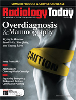
Overdiagnosis & Mammography
By Kathy Hardy
Radiology Today
Vol. 13 No. 6 P. 24
A study finds overdiagnosis with increased screening, but there’s no way to determine which tumors do not need to be treated.
As radiologists, oncologists, patients, and advocates continue to wrestle with when mammography screening should begin, a new study suggests that with more views of breast tissue comes more potential overdiagnosis of breast cancer. The study, published in the Annals of Internal Medicine, concludes that mammography screening entails a substantial amount of overdiagnosis, which could lead women to undergo unnecessary and potentially harmful treatments.
Some believe the research casts more doubt on screening mammography, a modality still dealing with an identity crisis in the wake of the US Preventive Services Task Force’s 2009 recommendations suggesting that women at normal risk of breast cancer can reasonably delay mammography screening until they reach the age of 50. However many people in the breast imaging field still recommend mammograms for women at normal risk begin at age 40. The split creates a decision for referring physicians and patients regarding when to start breast cancer screening. Many breast radiologists contend that the idea of overdiagnosis and potentially unnecessary treatment of nonfatal cancer adds to the dilemma for doctors.
“This new epidemiological study tries to show that if we weren’t screening so much, we wouldn’t find as many unimportant tumors,” says Robin B. Shermis, MD, MPH, medical director of Ohio’s Toledo Hospital Breast Care Center. “This study deals in a theoretical world. In practicality, we can’t always tell which tumors have a potential aggressive biology when they are first detected. At initial detection, there is no way to identify whether or not a tumor is life threatening or will become life threatening.”
In blunter words, if you can’t differentiate between the tumors that will progress and kill a woman and the ones that will never harm her, how do you decide which tumors to treat? Breast radiologists assert that it’s too early to discuss what to do when mammography uncovers a tumor that fulfills the laboratory criteria of cancer but, if left alone, would never cause the patient any harm. They contend that, since science cannot accurately predict which tumors are harmless and which are more aggressive, it’s necessary to treat any tumor that's found as if it's deadly. That means surgical removal and sometimes radiation or chemotherapy.
“That’s exactly the problem,” says Rulla M. Tamimi, ScD, an associate professor of medicine at Harvard Medical School and a coauthor of the study. “Through imaging and pathology, we’re unable to determine the difference between fatal and nonfatal cancers. It’s important to have studies like this to get the debate going. Women should know about overdiagnosis.”
Finding Too Much?
The objective of the report, “Overdiagnosis of Invasive Breast Cancer Due to Mammography Screening: Results From the Norwegian Screening Program,” was to estimate the percentage of overdiagnosis of breast cancer attributable to mammography screening. This was done with a comparison of invasive breast cancer incidence with and without screening.
Tamimi says the data from Norway provided a unique opportunity to review data collected during the county-by-county introduction of a breast cancer screening program for women aged 50 to 69 that took place from 1996 to 2005. Researchers analyzed approximately 40,000 breast cancers, comparing cases found in counties where screenings were offered against counties where screenings were not yet offered. The study’s authors found that instances of invasive breast cancer increased 18% to 25% among participants who received screening mammography. They also found that between 1,169 and 1,948 of those women were overdiagnosed and received unnecessary treatments.
“In any screening program, there will be risks and benefits,” Tamimi says. “One of those risks is detecting cancers that, if left alone, will not cause mortality in the population of people screened.”
Carol H. Lee, MD, FACR, attending radiologist at Memorial Sloan-Kettering Cancer Center in New York and chair of the ACR’s Breast Imaging Commission Communications Committee, notes that the Norwegian study findings agree with those of the US Preventive Services Task Force recommendations, which suggested that there is a risk of overdiagnosis based on the number of women screened. However, she’s not suggesting that this means cancers found in breast tissue should be left alone.
“Saying [there is overdiagnosis], I know that screening with mammography saves lives,” Lee says. “The emphasis on overdiagnosis is too great.”
Lee contends that screening mammography shouldn’t decrease just because it may find cancers that are not deadly. “Does it make sense to stop finding cancers because some of them will not go on to be fatal?” she asks. Lee also challenges the use of the term “overdiagnosis,” saying instead that nonlethal breast cancer may be overtreated, not overdiagnosed.
“If a tumor meets the histologic criteria for being malignant, we treat them all as if they’re life threatening,” Lee says. “That’s the trade-off. We can’t tell whether it is life threatening.”
Treat What You Find
Looking at the study’s parameters, radiologist Stamatia Destounis, MD, managing partner of Elizabeth Wende Breast Care in Rochester, New York, questions the validity of the data, noting that “if you want to prove something invalid you can look at the data any way you want.” In particular, she points to the time span used to gather data for this study.
“With mammography, you need to study a program for more than nine years,” she says. “Imaging to detect breast cancer involves identification of subtle findings on mammography over time and long-term follow-up after breast cancer diagnosis to identify long-term benefits. We need more information on the women within the study and the control group over longer periods of time to identify a benefit.”
In her work with the ACR, Lee spends a great deal of time discussing the benefits of breast cancer screening. With the publicity that surrounds studies like this and the task force recommendations, she says referring physicians and women are unsure of what steps they should take when it comes to mammography.
“Another benefit of screening mammography relates to treatment options,” Lee says. “If you have a mammogram, there’s a chance that it will pick up a cancer that will never be life threatening, but you’ll still undergo surgery and possibly chemotherapy and/or radiation. However, there is also a chance that your life will be saved and treatment for the cancer will be less invasive because it is caught sooner. Which would you prefer? Different women will have different reactions.”
One of Lee’s arguments against studies like this and the task force recommendations is that with an epidemiological study you’re dealing with mathematical modeling rather than with actual practice. Shermis agrees that epidemiology has its place when studying breast cancer from a public health perspective. However, while the studies may provide insight to cost factors associated with unnecessary medical procedures and the stress associated with screening mammography follow-ups, they don’t address the human aspect of detecting the disease in women.
“Yes, there’s anxiety associated with any follow-up related to screening mammography findings, but that’s minimal compared to knowing you have a tumor and not treating it,” Shermis says. “An option would never be to leave a cancer alone. In addition, there’s much less stress involved and it’s much less expensive to treat a cancer when it’s small than after it has grown large.”
Confusing Referrers and Patients
Shermis and others believe these study results simply add to the already confusing amount of information disseminated in the past several years regarding screening mammography guidelines.
“This is just more misleading info,” Shermis says. “Women and referring physicians are confused enough. We were just starting to see a bounce back in screening mammography from the backlash that resulted when the task force findings were issued. Now, this study compounds the confusion.”
Identifying cancer is not a perfect science, Shermis notes. However, it is the job of breast imagers, oncologists, and surgeons to follow the proper steps required to make the best educated decision possible when it comes to breast cancer detection and ultimately a course of treatment.
“Nothing’s perfect,” he says, “but when you have the right people involved, it’s a relatively smooth process. Until we can identify cancers, we need to treat tumors that we find. Breast cancer screening has been profoundly successful in saving lives. As long as you have good standards for how to work up cancers and you follow them, you will have success.”
Radiologists, surgeons, and oncologists “all recognize that this tumor may not do anything,” Shermis adds. “None of us could look at a cancer and say we didn’t have to treat it. However, we’ve seen tiny cancers metastasize and large cancers do nothing. We’re not in a position to guess whether or not a tumor could lead to cancer.”
“The continuing dilemma for breast imagers is that we try to be as evidence-based as possible,” Lee says. “We’re not resting on our laurels. Clinical trials done with a half-million women over more than 20 years show us that mammography is still the gold standard for breast cancer screening. We need to stop picking apart the basic finding that mammography saves lives.”
Identifying Dangerous Tumors
Rather than a continued focus on the ethicality of breast cancer screening, Lee suggests that researchers look to finding a way to sort out which cancers have the potential to be lethal and which are safe to leave alone. Tamimi believes that’s where data from the Norwegian study can actually be used to help the evolution of breast screening guidelines. Pointing out instances of overdiagnosis and overtreatment of tumors found in the breast can help researchers determine where to focus next in the process of developing accurate breast cancer detection methods. Advancements in imaging technology and the use of ultrasound and MRI in scanning breast tissue continue to help locate tumors but, in many cases, also increase the incidence of false-positives. But there are other areas where further research could help identify what the technology is finding.
“They need to look at tumor characteristics and tumor markers and learn more about which traits are less aggressive,” Tamimi says.
“Many women aren’t even aware that overdiagnosis exists in breast cancer screening,” she adds. “The discussion started with prostate cancer, but more should be said regarding overdiagnosis in breast cancer. Women are being told they have a cancer, which comes with its own stress, and then they have to deal with treatment. They should really have a clear picture of whether or not what was found in their breasts is good or bad before making those decisions.”
For the immediate future, however, Tamimi understands how these findings can create confusion for women over time. “It’s disappointing to people to hear that screening mammography isn’t the tool that it’s been presented as,” Tamimi says. “Dialogue about overdiagnosis is important to get out there so that research and developments don’t stay stagnant.”
Tamimi says the Norwegian study serves as a starting point for more effective communication between physicians and patients regarding overdiagnosis, which she contends goes hand in hand with misdiagnosis.
Lee points out that while this study brings up the issue of too much screening and the potential for overdiagnosis that might come with that, at the same time state and federal governments are debating the legislation of mandatory breast density notification. Texas, Connecticut, and Virginia passed laws within the last two years that require radiologists to notify patients if they have dense breast tissue following routine screening mammography; other states, as well as the federal government, are considering similar measures this year. There is the belief that legislating dense breast notification could lead to more screening with ultrasound, MRI, and other imaging modalities, which could lead to more false-positives.
“On the one hand we’re saying there are too many false-positives and too much screening,” Lee says. “On the other hand, there is the breast density notification issue that will likely lead to more screening. As breast imagers we’re caught between two imperatives: screen less vs. screen more.”
While Lee recognizes that mammography is not perfect, it is the only screening tool that has been shown to decrease mortality from breast cancer. “The bottom line is that screening mammography saves lives,” she says.
— Kathy Hardy is a freelance writer based in Phoenixville, Pennsylvania. She is a frequent contributor to Radiology Today.

