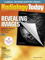
 Revealing Images
By Beth W. Orenstein
Radiology Today
Vol. 18 No. 10 P. 12
AI tools help radiologists look for cancers in dense breasts.
About 40% to 50% of women aged 40 to 74 in the United States have dense breast tissue, according to the Susan G. Komen foundation. Dense breasts are predominantly composed of tissue compared with nondense breasts, which are predominantly composed of fat. While not abnormal, dense breasts are linked to a higher risk of cancer. Very dense tissue, much like bone, shows up as white on a mammogram. Cancer, too, is white.
"When you're reading a mammogram, you're looking for white on a white background," says Christine Podilchuk, CEO of Koios Medical in Piscataway, New Jersey. "In these women, finding cancer is like looking for a polar bear in a snowstorm."
Women with dense breasts are often advised to have digital breast tomosynthesis (DBT) rather than plain 2D screening mammography. DBT has been shown to have a higher cancer detection rate, finding 29% more cancers and 41% more invasive cancers than 2D mammography, while reducing false-positives up to 37%, according to iCAD of Nashua, New Hampshire. The problem with DBT, however, is that it generates a vast amount of data. Conventional 2D mammography generates four images that a radiologist has to read and interpret; the same study done by DBT can generate hundreds of images.
More than 48 million mammograms are performed annually in the United States, according to the Centers for Disease Control and Prevention. According to the National Cancer Institute, more than 50% of women screened annually for 10 years in the United States will experience a false-positive result. When mammograms fall into the "suspicious" category, which could mean anywhere from 3% to 95% cancer risk, patients are typically advised to undergo a breast biopsy. According to the American Cancer Society, more than 1.6 million breast biopsies are performed annually in the United States. Of those, the Society estimates that about 20% are due to false-positive mammogram results.
Often, when mammogram findings are suspicious, women, especially those with breast dense tissue, are sent for a follow-up ultrasound. "The good thing about ultrasound," Podilchuk says, "is that it does find lesions in dense breast tissue, and the lesions it finds are the kinds of cancers you want to treat, those that are invasive but node negative; they have yet to spread to the nodes," she says. Another advantage of ultrasound, Podilchuk notes, is that it is low cost compared with other imaging technologies such as MRI.
Reading Glasses
In both cases, with DBT and ultrasound, artificial intelligence (AI) may become a key tool for improving breast cancer screening and diagnosis. Koios Medical recently introduced an AI platform to help radiologists distinguish breast cancer from benign/nonthreatening conditions on ultrasound, effectively reducing unnecessary biopsies and misdiagnosis. The tool, Koios DS, uses machine learning algorithms to assist radiologists as they interpret diagnostic images.
"It's like a pair of prescription eyeglasses—increasing the effectiveness of radiologists and reducing the need for biopsies," Podilchuk says.
"All radiologists are different," says Richard Mammone, PhD, founder of Koios Medical. The number of false-positives varies depending on who is interpreting the ultrasound, Mammone says. "Our system allows computers to make recommendations for radiologists based on tacit information. Tacit information is information you can't translate easily."
The system looks at thousands of breast ultrasound cases that have been read and, from them, determines which features are most likely associated with cancer and which are not, Mammone says. "We've trained neural networks to discriminate between subtle visual differences in cancers and harmless conditions. The software will assist radiologists in determining what is highly suspicious and should go for biopsy and what is not."
In today's health care environment, radiologists reading breast mammography and ultrasound are often faced with significant time constraints, Mammone says. "They have to make a lot of decisions quickly and accurately." The Koios DS software provides AI that reduces the cognitive load on radiologists, he says. AI is more advanced than previous computer-aided detection (CAD), which has been around for decades, he adds. Mammone says it can detect patterns that radiologists might miss and can determine, with more certainty, whether a lesion is malignant or not.
The National Institutes of Health funded a study of Koios DS, which was done in collaboration between Podilchuk, Mammone, principal investigator Susan Love, MD, MBA, of the Dr. Susan Love Research Foundation of Encino, California, and Wendie Berg, PhD, FACR, of the University of Pittsburgh Medical Center, Magee Womens Hospital. The study was conducted at the University of Southern California and UCLA. In the study, Koios' AI platform was able to accurately identify breast cancers while reducing the number of benign breast lesions sent to biopsy—as recommended by board-certified radiologists—by 70%. In January of this year, the FDA granted 510(k) clearance to Koios DS software. It is being further evaluated by radiology groups in the United States.
"We are taking the knowledge of experts with many years of experience and transferring that knowledge to other people—radiologists, nurses, and other health care providers in the field," Mammone says.
Because ultrasound is low cost and easily portable, Podilchuk believes Koios' AI software could have great potential in parts of the world where women don't have access to breast cancer screening, let alone quality breast screening. In places where there are shortages of radiologists, women could, in the near future, go for breast cancer diagnosis or screening with ultrasound, perhaps even on their smartphones, Podilchuk says, and AI could be used to interpret the results. Although Koios' initial focus is on breast cancer diagnosis, the company plans to expand its AI platform to other diseases and imaging modalities, Mammone says.
Enhanced 2D
iCAD's PowerLook Tomo Detection applies AI to DBT, which has also been proven to be a more accurate screening test for women with dense breast tissue. DBT continues to gain momentum and shows great promise as more insurers are willing to pay for it. PowerLook scans every plane of the hundreds of DBT-generated images for regions of interest. If a region of interest is found, it is lifted from the plane and blended naturally onto a synthetic 2D image, without obscuring it with physical marks. Each region is linked directly to the plane where it was detected to provide radiologists with a navigation tool. The tool is designed to be used concurrently throughout the reading session.
"Every patient is unique, and cancers have many different appearances. When reading the scans, it's a very subjective call," says Bruce Schroeder, MD, medical director of Carolina Breast Imaging Specialists in Greenville and Wilson, North Carolina. "The image I see [with PowerLook] looks like a 2D image so we can get a very quick overview. It's like reading a superenhanced 2D view."
That the PowerLook image the radiologist views in 2D is helpful for comparison, Schroeder says. "When reading a scan, it's important to compare it to all the images you had before. That gives you a very good idea of what's changing. If you see patients every year or every other year, it's very important to compare this year's 3D with last year's 2D. If you have hundreds of slices compared to four, that is more difficult and time consuming."
Like Mammone, Schroeder says time management is critical for radiologists. "If, because of the enormous number of images, it took us 200 times longer to read each 3D case vs a 2D case, we'd do one case a day and be out of business," he says. "We have to manage our time, which is the most expensive thing we have in our business."
Also, Schroeder says, real estate is limited—only so many slices can be up on the screen at one time. PowerLook allows radiologists reading DBT to focus on the possible abnormalities. "It's a real timesaver on top of being more accurate and sensitive."
Schroeder says it takes some time to get used to working with PowerLook, which was FDA approved in March 2017. "There's a learning curve, as there is with everything," he says. "In the beginning you trust nobody, whether it's FDA approved or not.
"This AI software solution is very different from 2D CAD and what we are used to," Schroeder says. "After spending some time using the product, I find it's very good at detecting cancer and extremely helpful because it highlights important areas."
Radiologists need more help with women who have dense breasts, Schroeder says. That's where he thinks PowerLook may be most helpful. "PowerLook seems to mark a few things on every case, and those are the places I will spend my time, but they are not necessarily all positive findings."
By the Numbers
PowerLook has a density assessment algorithm that factors in multiple image characteristics including volume of dense tissue. PowerLook Density Assessment also looks at structure, texture, and fibroglandular tissue dispersion throughout the breast, which more closely aligns with how radiologists assess density scores, based on the ACR's BI-RADS, fifth edition. By delivering an automated, fast, and reproducible assessment, PowerLook Density Assessment can help identify patients with dense breast tissue who may benefit from supplemental screening, says Adrea Bennett, RT (R)(M)(BS), Center for Women supervisor at SwedishAmerican Hospital in Rockford, Illinois.
In the breast density world, radiologists often teeter between a B and C breast density score, for example, Bennett says. Radiologists often have to estimate based on look alone, without having any numeric value associated with the density. PowerLook generates a numeric density value.
"For radiologists, PowerLook Density Assessment makes reading breast density easier and is one less thing they have to worry about in their extremely busy day, which allows patients to get faster results," Bennett says.
Shandong Wu, PhD, an assistant professor of radiology, biomedical informatics, and bioengineering in imaging research in the department of radiology at the University of Pittsburgh, says AI is more helpful for interpreting breast scans than traditional CAD, which is based on predefined features or rules. "For example, if the shape or margins of a lesion is such and such, it's likely malignant or more likely benign," Wu says. "If you want the model to work well, you have to come up with really good features and be able to formulate them to the models."
AI doesn't require researchers to predefine any features as likely or less likely to be malignant, Wu says. It is based on labeled data—AI learns rules or features automatically from the labeled data fed to the neural networks, and the more data it sees, the better it gets at determining likely malignancies or identifying regions as benign.
"A large labeled data set is key for the accuracy/intelligence of AI because it essentially learns knowledge from this large queue of data," Wu says. He adds that AI can reduce the likelihood of missing an important finding in dense breasts because it can see features that the human eye may not.
Smart Assistants
Some people, mostly those on the technical side of AI, have said radiologists will be replaced by it in five to 10 years. Should radiologists be concerned that they will be replaced by machines that can read breast exams?
"I don't see it that way," Wu says. "I believe AI will surely replace part of the clinical imaging reading work that radiologists currently do, but it is not likely to replace radiologists."
Rather, Wu says, AI will make the radiologist's job more efficient and valuable. Instead of spending significant time sorting through and reading thousands of images, radiologists will be able to preidentify suspicious/tricky cases or regions of interest that require further attention and study with the help of accurate, reliable, and thoroughly validated AI.
"AI can do a lot of reading work, and, combined with the radiologist's knowledge about logic and reasoning, together they can do a better job," Wu says. "AI is a tool for a part of clinical imaging, but you would still need a physician to participate in the clinical workflow."
Also, Wu says, if AI makes radiologists more efficient, they can spend time on more valuable work, such as interaction with patients, participation in treatment effect evaluation, and integrative research of imaging data and other sources of clinical data. The future roles/working mode of radiologists, however, will be significantly affected by AI, Wu predicts.
"Radiologists should start paying attention to AI and its quick growth in clinical informatics," Wu says. "In the future, they may have to learn how to live and work together with a smart AI assistant in their daily practice."
— Beth W. Orenstein, of Northampton, Pennsylvania, is a freelance medical writer and regular contributor to Radiology Today.

