 Editor’s Note: Evolving Imaging Paradigms
By David Yeager
Radiology Today
Vol. 26 No. 6 P. 4
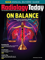
Even as medical imaging becomes more important in medical care, its modes and protocols for use are changing. Look no further than recent news for examples. There has been some controversy about the role that CT scan radiation plays in the development of new cancers. In April, a paper in JAMA Internal Medicine concluded that CT scans could be responsible for as much as 5% of cancers diagnosed in the United States in a single year. Professional societies such as the ACR and the American Association of Physicists in Medicine were quick to respond, claiming that the study’s findings were misleading.
In this month’s cover feature, Beth W. Orenstein provides an in-depth assessment of the disagreements that the study spawned, as well as some of the nuances in the respective arguments. Although people may disagree about the study’s findings, there seems to be broad agreement about the role of CT in modern health care: Everyone who was interviewed for the article agreed that CT should be used when necessary, and only when necessary.
The appropriate use of CT, as well as MR, is particularly relevant to the diagnosis and treatment of traumatic brain injuries (TBIs). Jessica Zimmer reports on the latest protocols for assessing TBI, which go beyond imaging and the use of the Glasgow Coma Scale. Recent changes in assessment incorporate additional data, such as blood biomarkers and a patient’s medical history, to develop a more complete understanding of a patient’s condition. In addition, technological developments, such as mobile imaging, have the potential to speed up assessment and diagnosis.
In other news, Keith Loria has an update on the state of the radioisotope market. Demand for radiopharmaceuticals has never been higher, and companies are making efforts to satisfy it. As PET grows to encompass theranostic treatments, demand will almost certainly increase further. Turn to page 18 for more on how this market is shaping up.
Finally, Orenstein has the details on a modality that’s part MRI and part MR spectroscopy. MR spectroscopic imaging (MRSI) incorporates aspects of both techniques, allowing researchers to see not only structural and functional information in the brain but also metabolic changes. Earlier versions of the technique were time intensive, but new, faster methods are making MRSI more useful for neurological imaging, potentially allowing clinicians to spot disease before symptoms appear.
Enjoy the issue.
— Dave Yeager
david.yeager@gvpub.com

