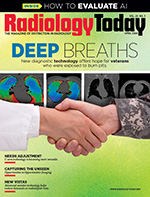
 Editor’s Note: Yet to Be Seen
By David Yeager
Radiology Today
Vol. 25 No. 3 P. 4
Seeing things in a new way has been the heart of medical imaging since Wilhelm Roentgen discovered X-rays. More than a century later, the field of radiology has advanced in ways that Roentgen would marvel at, but there is still more that is possible. In this issue, we’re looking at ways that imaging has revealed or enhanced physicians’ understanding of the human body, as well as ways that technology makes their job more efficient and improves patient care.
Our cover feature highlights veterans’ health. Many veterans who were deployed in Iraq and Afghanistan were exposed to burn pits. In some cases, the fumes from these pits caused deployment-related respiratory diseases (DRRDs), but until recently, the only way to positively identify many of these conditions was through lung biopsy. Claudia Stahl reports on imaging advances that now allow physicians to see DRRDs without resorting to biopsies. These technologies are changing the way veterans’ lung conditions are identified and managed.
Rebecca Montz also has a report on technologies that reveal previously unseen conditions. Medical scans contain a wealth of information, but only some of it is accessible. Gathering and analyzing more of this data offers a faster, less expensive opportunity to learn more about a patient’s overall health, not to mention ease the burden of multiple scans for patients who require frequent imaging. For exams that use ionizing radiation, such as X-ray and CT, this practice has the added benefit of reducing patients’ lifetime radiation exposure. Although opportunistic imaging isn’t a new concept, efforts to put it into practice are gaining momentum.
Doing more with less has long been a focus of radiology departments, and Keith Loria looks at how that trend is being incorporated into C-arms. As more and smaller facilities look to make use of C-arms, the equipment is getting smaller and adding features. AI, 3D imaging, image enhancements that produce quality images at lower radiation doses, and, in some cases, portability are being incorporated into these versatile machines.
Another piece of equipment that is rapidly evolving is the medical monitor. Beth W. Orenstein has a round-up of the latest features. Enhanced graphics, flexible configurations, and AI are all adding value to these essential tools. New monitors also help users customize workflows, an especially important consideration now that many radiologists read remotely. In addition, some monitors can identify and quantify dense breast tissue on mammograms, helping clinicians better diagnose breast cancer. For all of the details, turn to page 20. Enjoy the issue.
Enjoy the issue.
— Dave Yeager

