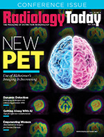
 New PET
By Beth W. Orenstein
Radiology Today
Vol. 26 No. 4 P. 10
Use of Alzheimer’s Imaging Is Increasing
A new study published in Nature Medicine in January 2025 predicts that by 2060, the number of people in the United States living with dementia will nearly double. That would mean one million new cases a year of the disease that involves progressive declines in memory, concentration, and judgment. The most common form of dementia is Alzheimer’s disease, which accounts for 60% to 70% of all cases, according to the World Health Organization (WHO).
Dementia is the seventh leading cause of death and a major cause of disability and dependency of people worldwide, WHO says. In 2019, the latest year for which statistics are available, dementia costs were about $1.3 trillion a year, mostly for caregiving, WHO says.
The prediction that the number of people living with dementia is likely to significantly increase makes recent developments in Alzheimer’s brain imaging good news. PET imaging plays a critical role in identifying lesions associated with the diagnosis of Alzheimer’s disease. No one can say exactly what causes Alzheimer’s, but amyloid plaque, sticky protein aggregates found in the brain and other organs, has become a hallmark of the disease. At some point, amyloid buildup appears to trigger tau proteins to start killing neurons, which leads to cognitive decline.
In Alzheimer’s disease, tau tangles are correlated with cognitive decline. The more tangles, the more severe the brain’s decline. PET imaging allows physicians to stage Alzheimer’s disease by quantifying the amount of tau in brain tissue, which helps them to determine the best treatments.
Updated Criteria
At the end of 2024, the SNMMI and the Alzheimer’s Association updated appropriate use criteria (AUC) for amyloid and tau PET imaging in patients with mild cognitive impairment, Alzheimer’s disease, and other forms of dementia. The new criteria expand the use of PET to include tau PET, incorporate updated information, and provide specific guidance that makes dementia diagnoses more precise. The new criteria also are meant to help physicians optimize patient care.
The late 2024 update is the first revision since the initial AUC for amyloid PET was introduced more than 10 years ago—in 2013. In that time, the CMS has announced key changes to reimbursement that make advanced imaging more accessible. In 2023, CMS lifted its one-scan-perpatient restriction and, in 2024, unbundled payment for high-cost diagnostic radiopharmaceuticals. The updated AUC also introduces guidelines for tau PET imaging. The new tau PET imaging guidelines pave the way for its broader clinical use. The new guidelines follow the FDA’s approval of the tau radiotracer 18F-flortaucipir in 2020.
Kevin Donohoe, MD, chair of SNMMI’s Committee on Guidance Document Oversight, says that the new AUC criteria focus on “optimizing patient care.” In an SNMMI press release on the new AUC, Donohoe says: “They will help providers determine the most effective use of these important PET tracers as well as describe clinical scenarios that are not likely to benefit from PET imaging. The AUC also discuss the use of PET imaging for determining eligibility for newly introduced dementia treatments and for following treated patients for response to therapy. It is also expected these AUC will reduce the need for less specific diagnostic testing and provide guidance for safety considerations.”
In a related development, a study in the January issue of The Journal of Nuclear Medicine found that two new radiotracers have outperformed 18F-flortaucipir, the only current FDA-approved radiotracer for detecting tau tangles in the brain. The study tested three different tau radiotracers to determine how well they could discriminate late-stage Alzheimer’s disease brain tissue from healthy brain tissue. A significant difference in binding was noted between the Alzheimer’s disease brain tissue and the healthy brain tissue in the whole brain hemisphere (prefrontal cortex) and hippocampus, but not for the cerebellar cortex for all three radiotracers. Binding to Alzheimer’s disease brain tissue was higher for the new radiotracers, 18F-MK6240 and 18F-PI260, than for the FDA-approved 18F-floratuacipir. The two new PET radiotracers also had greater selectivity than 18F-flortaucipir.
The study authors believe that their higher specificity makes the new tau imaging agents ideal for detecting the small changes that occur in brain tissue over time. A study author, Eduardo R. Zimmer, PhD, an assistant professor of pharmacology at the Universidade Federal do Rio Grande do Sul in Porto Alegre, Brazil, expects the new tracers to be especially helpful in Alzheimer’s disease clinical trials that utilize tau PET as an outcome measure. Additionally, Zimmer says, the study indicates that harmonization methods are needed to circumvent the differences between the imaging agents. “Our work represents an important step toward the harmonization of tau tracers and the results might provide insights into initiatives to create a universal scale for tau tracers,” Zimmer says.
Sea Change
Several important developments have occurred since the initial AUC criteria for PET amyloid imaging were released in 2013. Three developments include FDA approval of two additional PET amyloid tracers, 18F-flutemetamol, and 18F-florbetaben; FDA approval of 18F-flortaucipir, the first tau tracer; and a huge US national trial showing how PET amyloid imaging can change the diagnosis, says SNMMI spokesman Jacob Dubroff, MD, PhD, an associate professor of radiology in the division of nuclear medicine imaging and therapy at Perelman School of Medicine at the University of Pennsylvania and chair of SNMMI’s Brain Imaging Outreach Working Group. A fourth important development is multiple antiamyloid therapy trials using PET amyloid imaging leading to the FDA approval of two presently available therapies, lecanemab and donanemab, Dubroff says.
An SNMMI spokesman, Phillip H. Kuo, MD, PhD, FACR, a professor, division chief of nuclear medicine, and director of theranostics at City of Hope National Medical Center in Duarte, California, and coauthor of the AUC, says FDA approval of therapeutic antiamyloid antibodies that slow the progression of Alzheimer’s disease is part of a sea change that has occurred since the release of the original AUC. As a result of these many changes, he says, “We now use PET to image the two pathologic proteins of Alzheimer’s disease, amyloid and tau.” Dubroff and Kuo agree that the AUC needed to be updated.
Kuo says the AUC changes are highly significant. “A huge change was not only updating the AUC for amyloid PET but also adding AUC for tau PET,” he says. Also, new clinical scenarios were added, such as guidelines for handling the new therapeutic antiamyloid antibodies.
Dubroff says the new guidelines are significantly more complex. Prior guidelines identified how a single biomarker (PET amyloid) could support dementia diagnosis. “The new guidelines outline how two PET biomarkers, amyloid, and tau, can support diagnosis and direct care, specifically antiamyloid therapy,” Dubroff says. In the 2013 guidelines, the merits of using PET amyloid imaging in 10 different scenarios were examined. In the new 2025 release, 17 scenarios were examined using two different PET biomarkers (amyloid and tau)—essentially 34 in total.
Emerging Treatments
The new guidelines were developed by a multidisciplinary panel made up of 16 experts with backgrounds in PET imaging and clinical evaluation of patients with suspected dementia. The panelists reviewed the most recent literature and identified 17 scenarios in the current landscape where PET amyloid or PET tau imaging might be considered, Dubroff says. They issued scores from one to nine for each of the scenarios based on their review of the medical literature, with one being the lowest and nine the highest.
In 2023 and 2024, the FDA approved two new Alzheimer’s treatments: Leqembi (lecanemab) and Kisunla (donanemab), both monoclonal antibodies that target amyloid plaques in the brain, aiming to slow the progression of the disease in its early stages.
Kuo says amyloid PET can be used to determine eligibility for this antiamyloid antibody therapy and whether the antiamyloid antibody therapy is effectively removing amyloid from the brain. Dubroff adds that the patient’s clinical scenario should help determine whether to begin this therapy. “If someone with cognitive decline as assessed by a clinical dementia expert is thought to have Alzheimer’s, PET amyloid imaging could help determine their eligibility for antiamyloid therapy,” he says.
PET imaging can also be used to help determine the effectiveness of treatment. “Antiamyloid therapy serial imaging after initiation of therapy can be used to monitor response to therapy,” Kuo says. “And if a patient converts to negative on an amyloid PET scan, the antiamyloid therapy can be stopped.”
Again, Dubroff adds, much depends on the clinical scenario. “For many of the scenarios, the literature supports just one PET amyloid scan,” he says. However, for one of the FDA-approved antiamyloid therapies, donanemab, PET amyloid imaging can be repeated to see if the amyloid has been eliminated from the brain, in which case the therapy should stop. “This could be the strategy for antiamyloid therapies moving forward,” Dubroff says.
Improved Patient Stratification
Kuo and Dubroff believe the Journal of Nuclear Medicine study on the new tau tracers also contributes significantly to the diagnosis and treatment of Alzheimer’s. The study identifies the “next generation” of tau PET tracers and these tracers have the potential to image tau at earlier stages, thus allowing more accurate, earlier diagnoses of Alzheimer’s. “This may also help with better stratification of patients for clinical trials and selection of therapy,” Kuo says.
Dubroff adds that it is always good to have more tools (radiotracers) to help physicians and scientists identify tau tangles. These two new compounds are considered second-generation tau radiotracers. “They were designed to have better binding to tau tangles and less off-target binding (nontau tangles),” Dubroff says.
The study in the Journal of Nuclear Medicine showed that these new radiotracers had stronger binding in slices of brain tissue. PET radiotracers are administered intravenously and need to circulate throughout the body, pass through the blood-brain barrier, and bind to their targets. “The next step here would be to compare PET imaging characteristics, and additional steps will be taken,” Dubroff says.
More tau tracers are in development. “With the integration of tau PET imaging into more clinical trials of Alzheimer’s disease, we are learning more about the prognostic and predictive ability of tau PET,” Kuo says.
While additional tau tracers are in development, it could take as much as a decade or more before they become available. “Developing a tracer and receiving FDA approval for it takes years,” Dubroff says. “It’s not for the faint of heart.”
Kuo says that, in addition to tau tracers, researchers need to be looking at agents for neuroinflammation, neuronal damage, and synaptic loss. “The more new tools that are available to inform us of disease pathophysiology, the earlier we can recognize disease and the better we can classify it,” Dubroff says. “In turn, this will lead to earlier interventions and better clinical outcomes.”
Forward Trajectory
Blood tests for diagnosing Alzheimer’s disease are also in development. These tests look for levels of amyloid and tau in the blood. Blood biomarkers could support diagnosis and treatment in many ways, Dubroff says; however, the relationship between blood biomarkers and the PET amyloid and PET tau imaging tests needs to be better understood. Kuo believes that, although the topic is complex, blood and imaging tests could work synergistically to help each other with equivocal results.
The reality is that cognitive impairment is highly complex and patients may have mixed pathologies, Kuo says. “This is a challenge for diagnosis and treatment,” he says. “Patients with amyloid in their brains may have Alzheimer’s disease or another significant copathology, such as alpha-synuclein, which could indicate Lewy body dementia.”
Dubroff says it is striking how much has changed in one decade regarding brain disorders. “PET imaging for diagnosis and potential treatment of patients with suspected dementia has dramatically altered the landscape in the past decade and shows no signs of slowing down,” he says.
The updated AUC for amyloid and tau PET are available on the websites of The Journal of Nuclear Medicine and SNMMI.
— Beth W. Orenstein of Northampton, Pennsylvania, is a freelance medical writer and regular contributor to Radiology Today.
SIDEBAR
Brain Mapping Technique Provides Key Insights Into AD
Neurodegenerative diseases such as Alzheimer’s don’t affect all parts of the brain equally—some regions are more vulnerable than others. But why? The answer likely lies in the types of cells found in those regions.
Researchers from the University of Texas at Arlington and the University of California, San Francisco have used a new brain mapping technique to create detailed, brainwide maps of different cell types and subtypes. “We can then compare these maps with places where tau—a protein that accumulates abnormally in Alzheimer’s—is building up,” says Pedro Maia, PhD, an assistant professor of mathematics at the University of Texas at Arlington.
By comparing cell-type maps with tau accumulation in Alzheimer’s mouse models, the researchers uncovered key patterns:
• Hippocampal glutamatergic neurons are particularly vulnerable to tau buildup.
• Cortical glutamatergic and GABAergic neurons seem to be more resistant.
• Oligodendrocytes, a type of brain cell that supports neurons, showed the strongest negative association with tau accumulation.
Interestingly, Maia says, “We found that a brain region’s cell composition is a better predictor of tau buildup than the presence of known Alzheimer’s risk genes. This finding suggests that certain brain cell types are inherently more prone to damage in Alzheimer’s, offering new insights into how the disease progresses.”
This work also provides a better understanding of why some brain areas are hit harder than others, “bringing us one step closer to identifying potential therapeutic targets,” Maia says. The findings were published in February in the journal Communications Biology.
The brain mapping technique the researchers used is MISS—short for Matrix Inversion and Subset Selection. Published in PNAS in 2022, MISS is a computational method that combines these two datasets to estimate the spatial distribution of each cell type across the entire brain. In short, MISS helps create a detailed brain cell map by mathematically piecing together clues from different types of genetic data, Maia says. The researchers used MISS to profile about 1.3 million cells in the brains of mice.
To learn more about Maia’s work, visit the National Science Foundation Research Training Group Training in Mathematics for Human Health program, which he co-leads on computational neurology.
—BWO

