 MRI Monitor: A Quick Look
By Roni Shreter, MD, and Gil Shetrit, MBA, BSC
Radiology Today
Vol. 24 No. 8 P. 348
A novel deep learning image enhancement algorithm allows substantial improvement of MRI image quality and scan time reduction, according to a multisite study presented at the American Society of Neuroradiology annual meeting in April 2023. Low-resolution (LR) MRI images obtained by short protocols and automatically postprocessed by an AI-assisted algorithm were found superior to high-resolution (HR) images acquired with routine protocols.
In the past few years, radiologists have been facing a constantly rising demand for their services. Furthermore, they have to contend with repeatedly diminishing reimbursement rates and a shortage of personnel, leading to inevitable burnout. Breakthroughs in medical imaging operations became paramount. A recent multisite study conducted in the United States, Brazil, and Israel has shed light upon a promising development that holds the potential to alleviate these challenges.
Reducing MRI scan times can improve cost effectiveness, but the images acquired are often not adequate for diagnosis. This trade-off must be resolved in order to improve MRI efficiency.
Our research set out to evaluate a novel deep learning image enhancement impact on MRI imaging. The study specifically aimed to assess the overall diagnostic sufficiency of LR MRI images when processed through a deep learning algorithm (iQMR by Medic Vision Imaging Solutions) in comparison with images acquired at a higher resolution from various MRI scanners at multiple sites.
Testing the Algorithm
A group of 51 subjects, aged between 19 and 88 years, took part in the study. A comprehensive dataset was meticulously compiled, consisting of 195 sequences, encompassing various body parts, including brain, c-spine, l-spine, t-spine, knee, hip, finger, foot, and long bone images. What makes this dataset particularly compelling is that it was gathered from 12 distinct clinical MRI scanners, ranging from 1.2 T to 3 T, and sourced from reputable vendors such as Philips, Hitachi, Siemens, and GE.
Each patient underwent MRI imaging using their respective clinical site’s routine HR protocol. Additionally, a faster variant was applied to compromise resolution, reducing it by 25% to 33%, specifically in the phase encoding direction. The resultant LR scans were then subjected to a novel convolutional neural network-based image enhancement algorithm.
This algorithm was rigorously trained on an extensive dataset comprising MRI images from scanners of various vendors, showcasing a range of clinical indications. Its purpose was to produce enhanced resolution (ER) images.
Nine experienced radiologists conducted independent, blinded reviews. These assessments covered various aspects, including diagnostic quality, spatial resolution, noise levels, artifact level, and contrast resolution. Two comparisons were made: HR vs ER and LR vs ER.
Positive Outcomes
The results, drawn from a comprehensive dataset of 975 reads, showed that ER images consistently outperformed both LR and HR images across a spectrum of metrics (See Figure 1). For example, the diagnostics quality mean score for ER images is 5.21, compared with 4.81 for HR images and 4.65 for LR images. In addition, the results present significance for all mentioned criteria (p<0.001) (See Figure 2).
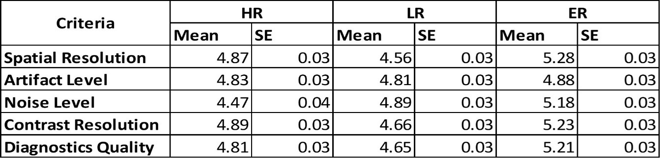
Figure 1: Descriptive statistics showing the overall ranking of ER images over LR and HR images in various criteria. Using a 7-point Likert scale (1=unacceptable, 7=excellent). Results demonstrate non-inferiority across all categories.
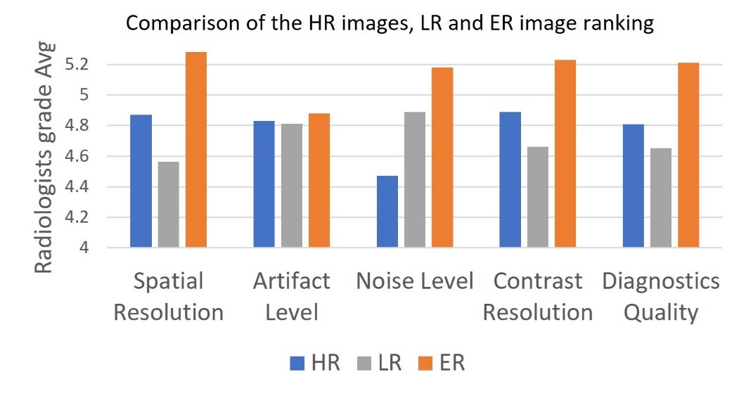
Figure 2: Comparison of the HR, LR, and ER image ranking.
Among the 158 reviewed series, 51 revealed pathologies, underscoring the clinical significance of this advancement and this technology’s potential to not only enhance image quality but also significantly contribute to the detection and characterization of clinically relevant conditions, further supporting its value in clinical practice (See Figure 3).
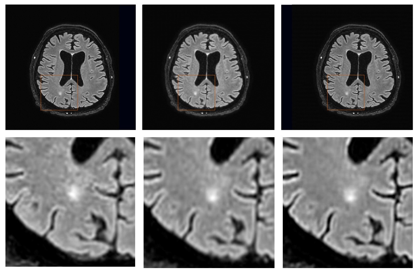
Figure 3: Comparing HR, LR and ER results. An example image of HR (left), LR (middle, 25% phase resolution reduction) and ER (right) of Axial PD FS images from a Philips Ingenia 1.5T scanner, showing a white matter hyperintensity clearer and more visible.
By harnessing the capabilities of a robust image enhancement AI algorithm, this study demonstrates the potential for substantial improvements in MRI image quality while simultaneously reducing scan times. This technological solution solves a longstanding trade off between resolution and acquisition time applicable to a spectrum of MRI scanners.
For radiology professionals, this development offers advantages in their day-today routine. As scan time is reduced, the workflow can be significantly improved, allowing for the exam schedule to be more efficient, accepting urgent cases, or relieving bottlenecks in patient throughput to answer constantly rising demand.
A shorter MRI scan may lead to another positive outcome: As the long wait inside an MRI bore often causes high levels of anxiety and discomfort, people may find it difficult to stay still. This, in turn, causes motion artifacts, which degrade image quality, and, often, result in the need for additional scans. With reduced scan times, patients enjoy shorter wait times, improving their overall health care experience.
— Roni Shreter, MD, is the director of the MRI unit in the diagnostic imaging department at Hillel Yafe medical center in Hadera, Israel.
— Gil Shetrit, MBA, BSC, is the CEO of Medic Vision Imaging Solutions.
