▼ ADVERTISEMENT

Editor's E-Note
Heart disease is the leading cause of death in the United States, killing approximately 695,000 people in 2021. In 2018 and 2019, it cost the United States $239.9 billion each year. Finding ways to better diagnose and track heart disease offers the possibility of saving lives and money. This month, we have a report from the United Kingdom on software that can create detailed images of the heart from MRI scans. The new technology creates precise 3D images of the heart valves and blood flow inside the heart, allowing physicians to diagnose and monitor heart disease more closely, potentially improving treatment planning.
Do you enjoy our newsletter? Let us know on Twitter and/or Facebook.
Enjoy the newsletter.
— Dave Yeager, editor |
|
|
| In This E-Newsletter
|
▼ ADVERTISEMENT
 |
|
|
Technology Detects Rapid Pressure Changes Inside the Heart
The state-of-the-art technology uses MRI to create detailed images of the heart. Using the new technology, researchers discovered that pressure inside the heart goes up when given a specific medication for testing the heart’s blood flow. They also found out why this medication, called adenosine, makes patients breathless during the test. The team says their findings could help doctors better diagnose and monitor patients with heart disease and heart failure.
Lead researcher Pankaj Garg, MD, from University of East Anglia’s (UEA) Norwich Medical School, says, “When patients present with symptoms of heart disease, doctors use a special test called heart MRI to take detailed pictures of the heart and see how well it is working. Sometimes, patients are given a special medication called adenosine during the heart MRI test to see how blood flows through the heart, and it can cause breathlessness. We wanted to better understand the way that the heart functions, and why patients become breathless when given adenosine.”
▼ ADVERTISEMENT
The UEA team worked with researchers at the University of Leeds and studied 33 patients referred for a stress cardiac MRI. This test is performed to help evaluate the blood flow in the heart arteries, looking for blockages. The research team took pictures of the patient’s heart when it was resting and when it was working hard after being given adenosine.
“Adenosine mimics the effect of exercise on the heart while the patient is lying down on the scanner,” Garg says, “And we discovered why it makes patients get out of breath.”
|
Study Demonstrates Effectiveness of MR-Guided Steering Strategies
A study published in Science Advances demonstrated the work of researchers utilizing a magnetic guidewire along with steering to increase the control and effectiveness of MRI. The technique could provide numerous intuitive ways for surgeons to use the technology.
Higher Muscle Fat Deposits Increase Mortality
Diagnosing asymptomatic adults with myosteatosis, or the accumulation of fat in muscle, poses difficulties. According to a study published in Radiology, these patients have an increased mortality rate, which makes diagnosis paramount.
Fluorescent Aid Detects Residual Tumor Post Breast Surgery
A fluorescent agent lit up at locations containing residual tumors following breast cancer surgery, according to a multicenter trial conducted by Mass General Cancer Center. |
“The use of large language models like ChatGPT is exploding and only going to increase. Our research provides insight into ChatGPT’s performance in a radiology context, highlighting the incredible potential of large language models, along with the current limitations that make it unreliable.”
— Rajesh Bhayana, MD, FRCPC, an abdominal radiologist and technology lead at University Medical Imaging Toronto, Toronto General Hospital, on news that ChatGPT passed the Radiology Board Exam and its future implications |
|
|
COVER STORY
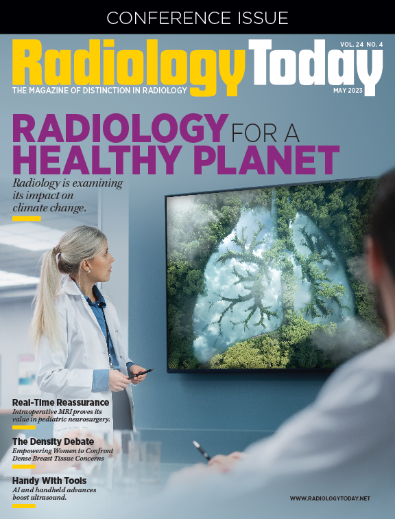
Radiology for a Healthy Planet
Radiologists are looking critically at the environmental impact of their practice and ways to mitigate advancing climate change.
FEATURE
Real-Time Reassurance
Intraoperative MRI is changing the field of pediatric neurological care, giving parents hope and patients better outcomes.
|
|
|
| Advertising Opportunities |
Have a product or service you want to market to radiology professionals? Utilize the reach of Radiology Today Magazine to accomplish your marketing goals. Email our experienced account executives today at sales@gvpub.com or call 800-278-4400 for more information.
|
| © 2023 Radiology Today Magazine |
|
|