|
E-Newsletter • March 2024 |
Editor's E-Note
Minimally invasive treatments to remove artery blockages are well established, but this month we’re highlighting a minimally invasive imaging technique that promises to improve visualization of arterial plaques. Although it’s not ready for use in humans, the research on its viability is progressing.
What minimally invasive procedures does your facility perform? Please let us know on X, formerly known as Twitter, and/or Facebook.
Enjoy the newsletter.
— Dave Yeager, editor |
|
|
| In This E-Newsletter
|
 |
|
|
New Approach Provides a Better View of Cardiovascular Plaques
Researchers have developed a new catheter-based device that combines two powerful optical techniques to image the dangerous plaques that can build up inside the arteries that supply blood to the heart. By providing new details about plaque, the device could help clinicians and researchers improve treatments for preventing heart attacks and strokes.
Atherosclerosis occurs when fats, cholesterol, and other substances accumulate on the artery walls, which can cause these vessels to become thick and stiff. A heart attack or stroke may occur if an atherosclerotic plaque inside the blood vessels ruptures or parts of it break off.
▼ ADVERTISEMENT

“Atherosclerosis, leading to heart attacks and strokes, is the number one cause of death in Western societies—exceeding all combined cancer types—and, therefore, a major public health issue,” says research team member leader Laura Marcu of University of California, Davis. “Better clinical management made possible by advanced intravascular imaging tools will benefit patients by providing more accurate information to help cardiologists tailor treatment or by supporting the development of new therapies.”
In the Optica Publishing Group journal Biomedical Optics Express, researchers describe their new flexible device, which combines fluorescence lifetime imaging (FLIM) and polarization-sensitive optical coherence tomography (PSOCT) to capture rich information about the composition, morphology, and microstructure of atherosclerotic plaques. The work was a collaborative project with Brett Bouma and Martin Villiger, experts in OCT from the Wellman Center for Photomedicine at Massachusetts General Hospital.
|
Researchers Find New Use for ‘Junk DNA’
Researchers from Johns Hopkins Kimmel Cancer Center have discovered a new use for the repeat DNA sequences frequently referred to as “junk DNA.” The discovery could lead to new, noninvasive methods of detecting cancer and monitoring receptiveness to treatment.
Emerging Technique Could Predict Heart Attack Risk
In a study published in Radiology, researchers found that utilizing coronary artery calcium scoring in conjunction with CT could potentially assist in identifying patients at varying risk of heart attack and stroke.
New Seizure Imaging Method Could Aid Brain Surgery
Research published in The Journal of Nuclear Medicine discloses a new way of inducing and imaging seizures. The data collected can be used to better inform the treatment for patients unresponsive to medication who require surgery to remove epileptic brain tissue. |
“Our results indicate nnU-Net's remarkable accuracy for 3D segmentation of prostate glands even with limited training data, offering a simpler and faster alternative to our previous 3D gland-segmentation methods. Notably, it maintains good performance with lower-resolution inputs, potentially reducing resource requirements.”
— Jonathan T. C. Liu, MS, PhD, a professor of mechanical engineering, bioengineering, and laboratory medicine and pathology at the University of Washington, and lead researcher on a study utilizing deep learning to improve prostate cancer detection and treatment |
|
|
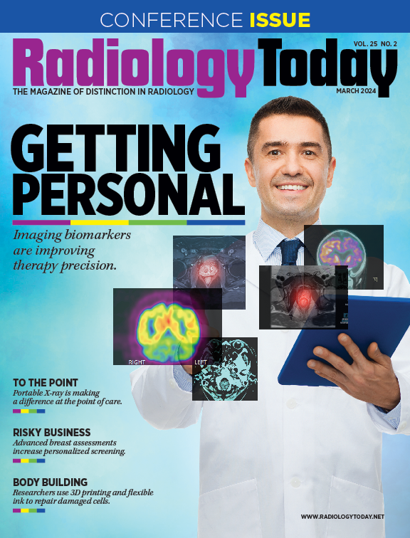
COVER STORY
Getting Personal
Every patient is unique; with the use of biomarkers and the aid of theranostics, imaging is getting more personal and precise than ever.
FEATURE
To the Point
Handheld X-ray at the point of care is making imaging more convenient and accessible.
|
|
|
| Advertising Opportunities |
Have a product or service you want to market to radiology professionals? Utilize the reach of Radiology Today Magazine to accomplish your marketing goals. Email our experienced account executives today at sales@gvpub.com or call 800-278-4400 for more information.
|
| © 2024 Radiology Today Magazine |
|
|