E-News Exclusive
Radiotherapy in Motion
 Traditional radiation therapy involves taking a reference image of a patient and planning treatment around it, but that method doesn’t account for movement of tumors, tissues, or organs. Movement is a critical challenge for radiotherapists. Tissues or organs that move may receive unintended radiation, and a tumor may not receive the prescribed dose.
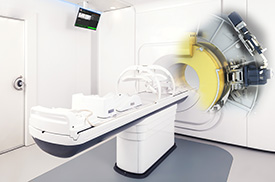
A newer method, online adaptive therapy, allows physicians to image a patient in real time and tailor radiation to the specific anatomy of the patient. The Unity system from Elekta combines a 1.5 T MRI scanner with a linear accelerator to precisely locate and track tumors. According to Kevin Brown, global vice president of scientific research for Elekta, treatment delivery can be “adapted in real time in response to changes in tumor position and shape and the relationship to sensitive organs over time. Due to the capability of the MRI, the system also offers the potential to respond to the biological characteristics of the tissue.”
Brown says the Unity system’s ability to provide up-to-date information allows clinicians to maintain smaller tumor margins when delivering treatment and deliver more dose to tumors while reducing dose to surrounding tissues and organs. Clinicians who are currently performing online adaptive therapy see significant potential for its use.
“MR-linac is a truly groundbreaking radiotherapy system that combines a modern, fully functional, diagnostic-quality MR scanner with an advanced linear accelerator. The ability to easily and consistently define the margin of a tumor will allow clinicians to tailor each radiotherapy treatment much more precisely than is currently possible,” says Christopher Schultz, MD, FASTRO, FACR, chair of the department of radiation oncology and Bernard and Miriam Peck Family Professor at the Froedtert & the Medical College of Wisconsin Cancer Network and chair of the Elekta MR-linac Consortium. “I’m also excited at the prospect of being able to manage tumor motion and actually monitor tumor regression during a course of radiation therapy. Such spatial and tumor response information will allow for even further refinement and personalization of each patient’s treatment.”
A study from the University of Utrecht Medical Center in the Netherlands published in November 2017 in Physics in Medicine & Biology looks at four patients with lumbar spinal bone metastases who were each treated with a single fraction of 8 Gy prescribed to the target volume, while the dose to the spinal cord and the remainder of the body was minimized. Quality assurance procedures were performed for the pretreatment and delivered plans, and imaging analyses were used to determine accuracy. The results found high dosing and positional accuracy while allowing treatment decisions to be made on the treatment table throughout radiation delivery, Brown says.
“I am excited about the prospect of personalized radiotherapy. For the first time, we will be able to truly intervene and adapt our radiotherapy based on an individual patient’s tumor response. This is not something that we have been able to do well with existing technologies,” says William Hall, MD, an assistant professor in the department of radiation oncology at the Froedtert & the Medical College of Wisconsin Clinical Cancer Center in Milwaukee. “My hope is that the integration of high-field MRI with a linear accelerator will truly enable personalized radiotherapy such that we can adapt care to meet the needs of individual patients each time they are treated.”
In June, the Unity system received the CE Mark, and the first patient was treated with it on August 22. It is pending FDA approval.
— A Radiology Today staff report
