


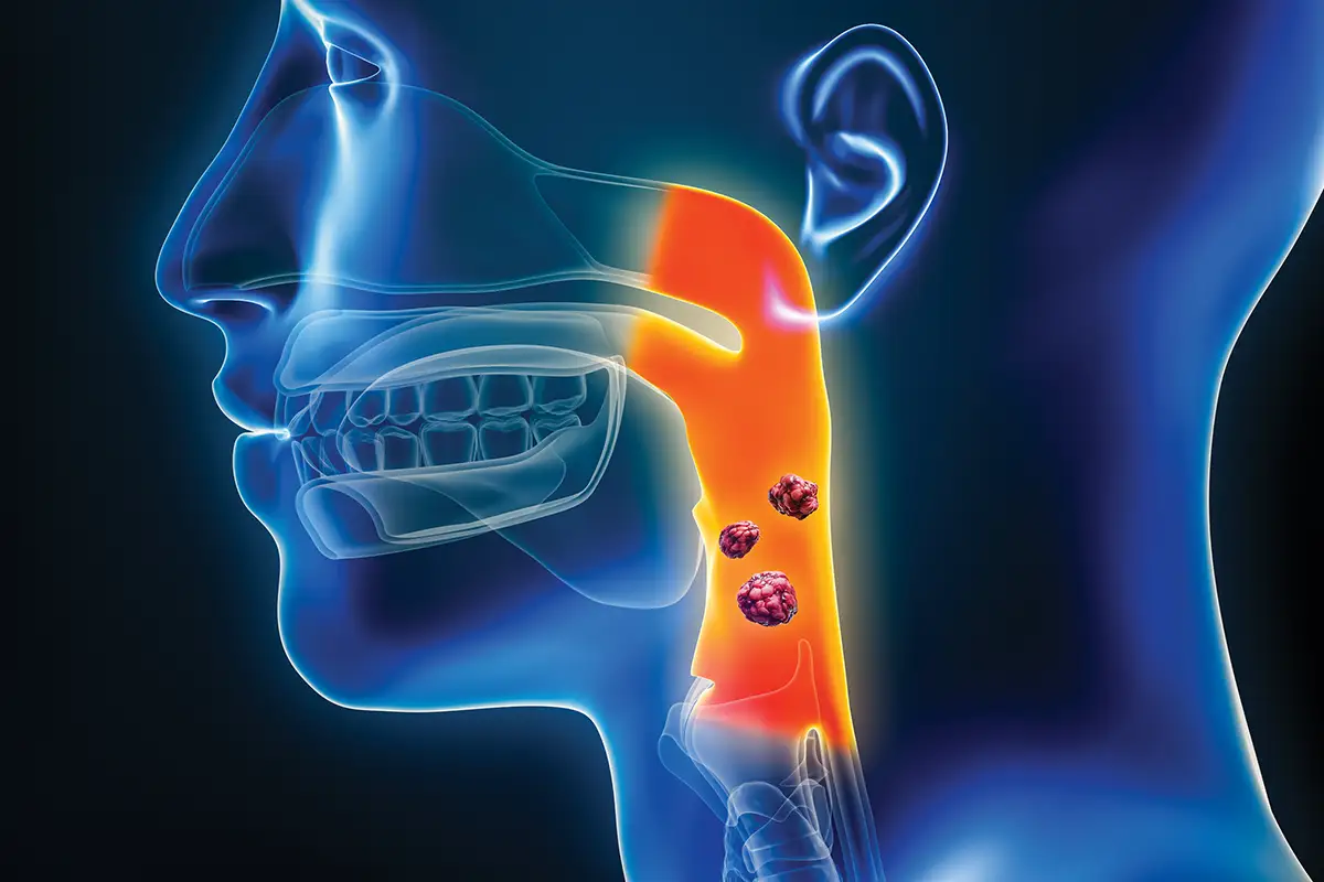
Segmentation of primary tumors on MRI is time-consuming and exhibits high inter-rater variability. There is immense potential for an automated algorithm that provides standardized segmentations and consistent measurements. While many AI segmentation algorithms for oropharyngeal squamous cell carcinoma (OPSCC) primary tumors have been developed to use both noncontrast and postcontrast sequences, the question of whether AI algorithms perform comparably on noncontrast vs contrast-enhanced sequences remains largely unanswered. In our recently published study in Diagnostics, we evaluated paired sequences from a 39-patient cohort at the University of Chicago to determine whether noncontrast T2 sequences alone could offer a viable baseline when contrast is contraindicated. Our analysis demonstrated that volumetric segmentation performance using noncontrast T2 sequences alone was statistically comparable to the combined, contrast-enhanced approach.
While contrast agents are undeniably useful for staging and identifying the spread of cancer, not all patients can safely receive them. Since contrast agents are processed by the kidneys, they carry a higher risk of nephrogenic systemic fibrosis in patients with renal impairment. Additionally, patients with documented allergies or concerns about retention in certain neural tissues may not safely receive contrast agents. In such situations, clinicians utilize noncontrast sequences or other alternative imaging modalities such as PET or CT to accurately stage and plan treatments.
OPSCC is one of the most common head and neck cancers, and HPV-positive cases have been increasing in incidence over the past two decades. During the pretreatment stage, clinicians often use MRI scans to help with staging and subsequent radiotherapy treatment planning. Pretreatment MRI studies typically include both precontrast and postcontrast sequences, which are acquired before and after injection of a contrast agent. In noncontrast sequences such as T2W, the tumor generally appears hyperintense, which helps the radiologist delineate the size and borders of the primary lesions. In postcontrast sequences, such as contrast-enhanced T1W, the tumor appears markedly bright (enhanced) relative to the surrounding healthy tissue, and these sequences also highlight the central necrosis within the tumor, which is especially common in HPV-positive OPSCC.
Comparing Three Sequence Configurations
The work was led by senior author Daniel T. Ginat, MD, MS, of the University of Chicago’s department of radiology. We used paired pretreatment MRI sequences (contrast-enhanced T1-weighted fat-suppressed and noncontrast T2-weighted fat-suppressed, acquired in the same imaging session) from 39 HPV-positive OPSCC patients enrolled in the OPTIMA II Phase 2 trial between October 2017 and February 2020. The sequences were coregistered, and manual tumor segmentation was performed by a radiologist with >10 years of clinical experience. The segmentations were reviewed by a senior board-certified neuroradiologist with >17 years of experience to establish an accurate ground truth. Using paired sequences from the same patient and imaging session controlled for interpatient variability that would otherwise confound the comparison. For this study, we trained and evaluated three separate model configurations: contrast-enhanced T1-weighted fat-suppressed (CE-only), T2-weighted fat-suppressed (T2-only), and a combined CE + T2 approach.
The nnU-Net is a self-configuring deep learning segmentation framework developed by Isensee et al. One of the most important features of this framework is that it automatically configures the optimal preprocessing, architecture, and training settings. This is important because it removes the algorithm as a confounding factor, and the differences in segmentation performance are primarily attributable to the input data.
The primary metric used to evaluate segmentation performance was the Dice metric. Dice coefficient measures the geometric overlap between the ground truth and the predicted segmentations. It ranges from 0 to 1, where 1 indicates that the predicted segmentation perfectly overlaps the ground truth, and 0 indicates that it completely misses it. In published literature, the Dice scores of AI algorithms often range from 0.55 to 0.75. These moderate scores highlight the difficulty of segmenting tumors in a complex region around the oral cavity, where various tissue types are present.
To study the boundary accuracy, ie, how closely the predicted tumor boundary aligns with the ground truth boundary, we used Surface-Dice@2mm. Surface-Dice@2mm measures the percentage of the predicted boundary surface that lies within 2 mm of the reference boundary and correlates with the time radiologists spend manually correcting AI-predicted contours. Performance was estimated using nested crossvalidation with eight outer folds for performance estimation and five inner folds for model selection, to produce an unbiased estimate from the modest sample size.
Comparable Performance, With Caveats
Across all three configurations, segmentation performance was statistically comparable, with noncontrast T2 sequences alone reaching levels close to contrast-enhanced T1. The median Dice was 0.63 for CE + T2, 0.60 for T2-only, and 0.55 for CE-only, with no statistically significant differences between configurations. For context, expert-to-expert agreement on this task hovers around 0.60, so all three configurations are operating within the range of human reader variability. The median Surface-Dice@2mm was 0.62 for the combined configuration, 0.60 for CE-only, and 0.57 for T2-only. The small reversal between the two metrics is consistent with what each measures. Surface-Dice@2mm rewards boundary precision, where contrast enhancement helps, while volumetric Dice rewards overall regional overlap, where T2’s broader tumor visualization is competitive.
While quantitative scores can provide an accurate picture of overall segmentation performance and errors, they often miss important qualitative aspects that can only be identified by an experienced radiologist through visual qualitative analysis. For example, predicted segmentations may incorrectly include surrounding healthy tissues, vastly underestimate tumor volume, and include distant islands of false positives far from the ground-truth tumor regions. Moreover, smaller tumors (under 3 mL, gross tumor volume) are harder to segment in both noncontrast and contrast-enhanced sequences. We conducted a blinded reader study in which the segmentations were qualitatively rated by a radiologist with >10 years of clinical experience on a 4-point ordinal scale (0 = reject, 1 = acceptable with major edits, 2 = acceptable with minor edits, 3 = acceptable as-is). The AI segmentations were rejected if the predicted segments included any substantial errors unlikely to be corrected with minor or major edits in the clinical setting. Across the three configurations, acceptability ratings were statistically similar, with 35% to 38% rated acceptable as-is or with minor edits.
Toward Contrast-Sparing Options
This study does not claim that T2 segmentation outperforms CE, nor that contrast-enhanced sequences contribute nothing of value. In fact, while Dice scores showed no statistical difference on volumetric bulk overlap, the Surface-Dice@2mm showed slightly better boundary alignment when contrast-enhanced sequences were included. Despite moderate performance, T2-weighted and combined contrast-enhanced + T2 sequences show significant potential to assist with contouring workflows. However, given a single-center, 39-patient cohort, this study underscores the need for a larger multicenter dataset with improved strategies for small tumor detection. Translating such recommendations to clinical practice would still require prospective evaluations with predefined clinical endpoints, not just Dice scores.
This preliminary, single-center study suggests that AI-based segmentation of HPV-positive OPSCC tumors on noncontrast T2 MRI may be a viable accommodation for patients in whom contrast is contraindicated. Broader claims will require multicenter prospective validation, evaluation against radiation oncology endpoints, and improved handling of small tumors. The full study is open access in Diagnostics (https://doi.org/10.3390/diagnostics16050658), with the analysis code on GitHub (https://github.com/rbramkumar/opscc_segmentation).
— Ramkumar Rajabathar Babu Jai Shanker, MS, is a research collaborator in the department of radiology at the University of Chicago and cofounder of Rad-Lab.ai, a research-translation initiative focused on clinically deployable medical imaging AI. He serves as a peer reviewer for the American Journal of Neuroradiology, the Journal of Magnetic Resonance Imaging, and the Journal of Computer Assisted Tomography. He is cofirst author of the study described in this article.
Disclosures
This article is an original submission, has not been published elsewhere, and is not under consideration by any other publication. This research was partially funded by Guerbet. The sponsor had no role in the design of the study; in the collection, analyses, or interpretation of data; in the writing of the manuscript; or in the decision to publish the results. The author and senior author Daniel T. Ginat are cofounders of Rad-Lab.ai. This work was completed in part with resources provided by the University of Chicago Research Computing Center.
Resources
1. Zarovniaeva V, Babu Jai Shanker RR, Shetty A, Ginat DT. Deep learning (nnU-Net)-based segmentation of primary HPV-positive OPSCC: contrast-enhanced T1-weighted fat-suppressed versus non-contrast-enhanced T2-weighted fat-suppressed MRI (paired single-center study). Diagnostics (Basel). 2026;16(5):658.
2. Isensee F, Jaeger PF, Kohl SAA, Petersen J, Maier-Hein KH. nnU-Net: a self-configuring method for deep learning-based biomedical image segmentation. Nat Methods. 2021;18(2):203-211.
3. Kanda T, Ishii K, Kawaguchi H, Kitajima K, Takenaka D. High signal intensity in the dentate nucleus and globus pallidus on unenhanced T1-weighted MR images: relationship with increasing cumulative dose of a gadolinium-based contrast material. Radiology. 2014;270(3):834-841.
4. Update on FDA approach to safety issue of gadolinium retention after administration of gadolinium-based contrast agents. US Food and Drug Administration website. https://www.fda.gov/media/116492/download. Published September 20, 2018.
5. Rosenberg AJ, Agrawal N, Juloori A, et al. Neoadjuvant nivolumab plus chemotherapy followed by response-adaptive therapy for HPV+ oropharyngeal cancer: OPTIMA II phase 2 open-label nonrandomized controlled trial. JAMA Oncol. 2024;10(7):923-931.