


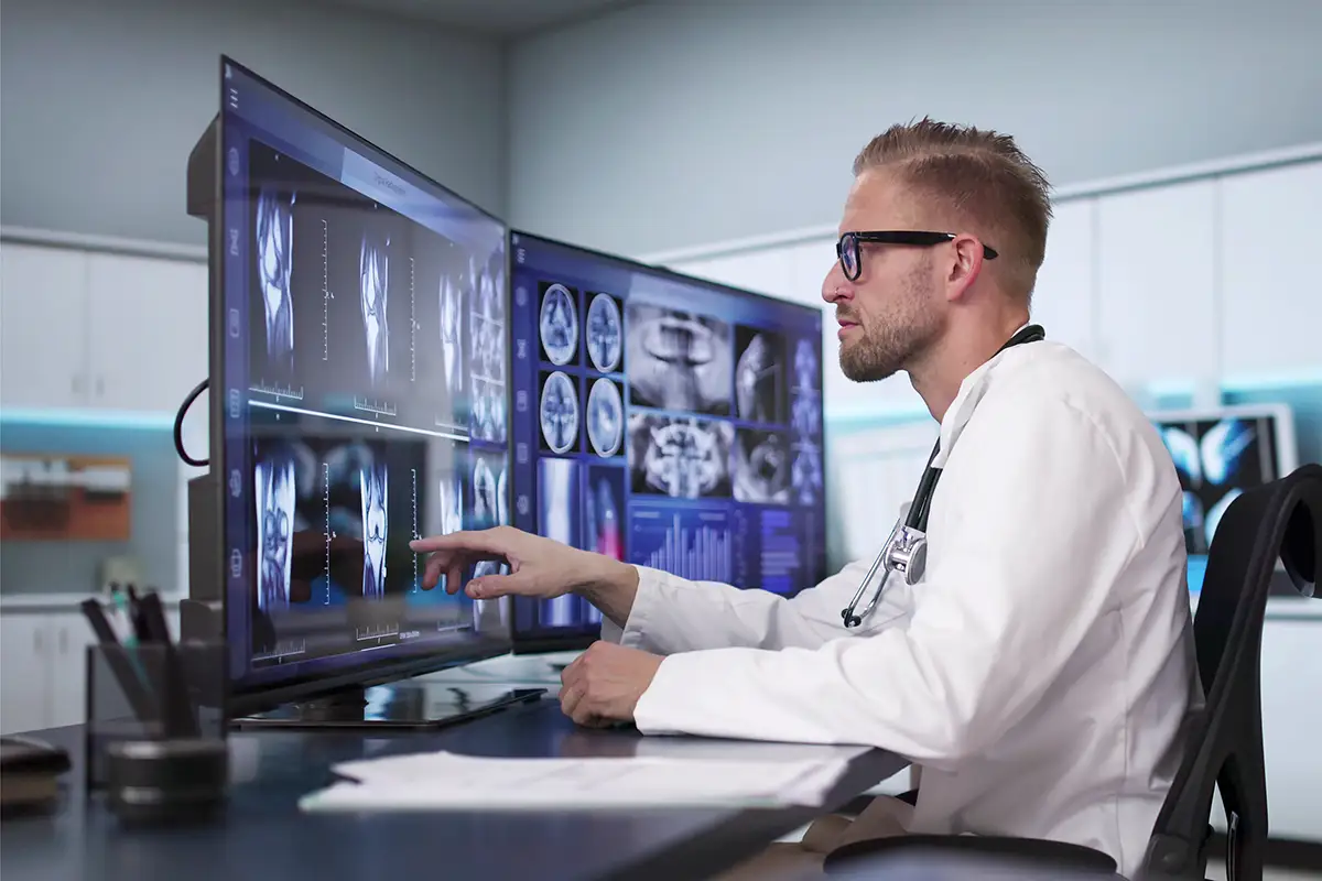
Historically, the diagnostic display has been evaluated through a familiar collection of specifications including resolution, luminance, contrast, uniformity, and grayscale performance. While those measures remain essential, they no longer tell the complete story.
As radiologists interpret growing numbers of studies from hospital reading rooms, outpatient centers, and home workstations, medical displays are becoming part of a larger image-management and workflow ecosystem. Manufacturers and display specialists are placing greater emphasis on centralized quality assurance, automated calibration, flexible configurations, ergonomics, and the ability to accommodate AI-generated findings without crowding the diagnostic image.
“The most significant advances are coming from the way displays with builtin front sensors, calibration software, and enterprise management tools now work together,” says Tara Neill, director of sales and marketing for Double Black Imaging. “Diagnostic displays are no longer viewed as standalone monitors. They are part of a complete reading environment.”
That shift has become particularly important as reading environments have become more distributed. A radiologist may interpret studies from multiple locations during the same week, but the expectations for image quality cannot change according to the workstation being used.
Neill notes organizations increasingly need tools that can monitor, test, and document display performance throughout the enterprise, including at-home reading stations. The issue has become especially relevant as remote breast imaging interpretation, or telemammography, expands.
Radiologists need stable luminance, consistent grayscale presentation, accurate color, and reliable quality assurance wherever they read, Neill says. Built-in sensors and calibration software help displays maintain that performance over time rather than relying solely on periodic manual checks.
Jim Salamon, senior health care IT manager at LG Electronics USA, also sees displays assuming a broader role in radiology operations. Health care organizations are focused on saving time, improving outcomes, and investing in technologies that contribute to operational performance, he says.
“Rather than being viewed as standalone equipment, displays are increasingly part of a broader workflow ecosystem designed to help radiologists work efficiently, maintain consistency, and trust what they see on screen,” Salamon says. The most effective solutions, he adds, support diagnostic confidence without introducing additional complexity into an already demanding reading environment.
One Workstation, Many Modalities
Although radiologists are reading a wider variety of studies from the same workstation, display requirements are also becoming more modality specific. As a result, for CT, MRI, digital radiography, and general radiology, DICOM calibration, grayscale accuracy, luminance stability, and image uniformity remain fundamental.
Mammography carries additional requirements because subtle differences in tissue appearance can influence interpretation. Breast imaging displays generally require higher resolution and luminance, along with the documentation needed to support quality control and regulatory compliance.
Digital pathology creates a different set of display demands. While diagnostic radiology has historically concentrated heavily on grayscale performance, pathology depends on reliable color reproduction, stain differentiation, brightness stability, and the ability to navigate extremely large, high-resolution, wholeslide images.
“In mammography, image quality and grayscale performance remain critical because even subtle details can influence a diagnosis,” Salamon says. “Digital pathology, on the other hand, places a premium on color accuracy and consistency.”
At the same time, the growth of multimodality reading means many users do not want separate workstations for every clinical application. Radiologists may move among CT, MRI, radiography, ultrasound, and other studies throughout the day. Displays increasingly must support that variety without sacrificing image quality or forcing users to repeatedly reconfigure the workspace.
Neill says flexibility has consequently become one of the most important purchasing considerations. Radiologists need room for current images, prior examinations, worklists, reporting applications, clinical information, and AI results. That demand is encouraging interest in larger-format diagnostic displays, integrated keyboard-video-mouse (KVM) functionality, and software that can manage multiple displays throughout a health care system.
Salamon says KVM switching can simplify a reading environment in which a radiologist must access more than one workstation or computing system. Instead of changing keyboards and mice or adding more hardware to the desktop, the user can control several connected systems from one set of peripherals. Ergonomics, reliability, and ease of use are also receiving closer scrutiny. A monitor’s technical image quality remains paramount, but departments are increasingly considering how the entire configuration affects radiologist efficiency and satisfaction over a long reading shift.
More Information on the Screen
AI is adding another layer of information to the display environment. Measurements, annotations, segmentations, probability scores, color-coded overlays, heat maps, and worklist prioritization alerts may all appear alongside the original medical image. That makes display accuracy more important.
“Displays must present that information clearly without compromising the original diagnostic image,” Neill says.
Many AI applications use color to identify suspected findings or distinguish anatomical structures. The display, therefore, needs to reproduce those colors predictably while preserving the grayscale characteristics and subtle image details needed for primary interpretation.
Screen space also matters. Radiologists need to view AI output in context rather than continually opening, closing, or moving windows. Larger and higher-resolution displays can allow the diagnostic image, priors, reports, and AI-generated information to coexist more naturally.
Salamon says displays are one of the primary points at which radiologists assess AI findings and decide whether to act on them. If the screen does not present that information clearly and consistently, confidence in the AI application may suffer. Displays are not performing the AI analysis, but their design can influence whether the results fit seamlessly into the reading process or become another source of distraction and window management.
“The key to successful AI adoption in health care will be centered on clinician trust,” he says. “Medical displays remain one of the primary touchpoints where radiologists validate and act on the information they’re given.”
Quality Across the Enterprise
Calibration and quality assurance have always been essential to diagnostic display performance. The expanding number and geographic distribution of workstations, however, are changing how departments manage those responsibilities.
“A medical display has to perform consistently, not just on the day it is installed, but throughout its life,” Neill says.
Manual inspection and one-time calibration are no longer enough for organizations managing displays across hospitals, imaging centers, clinics, and radiologists’ homes. Administrators need to know whether each monitor remains within the organization’s performance standards and whether required tests have been completed and documented.
Double Black Imaging’s CFS Calibration Software, for example, is designed to automate DICOM calibration, testing, reporting, alerts, and fleet management. Neill says centralized software gives PACS, IT, imaging, and compliance personnel greater visibility into display performance and can help identify problems before they affect interpretation or documentation.
The information being collected is also becoming more detailed. Rather than simply recording that a display passed a calibration check, health care organizations may want luminance histories, trend information, exception reports, test schedules, and records that can support internal quality programs, accreditation, or regulatory reviews.
Salamon says display management tools can help teams automate calibration and conformance testing, generate reports, and flag nonconformance issues. Continuous oversight provides a documented method of verifying performance rather than depending on the results of an installation-day test.
Centralized management may also reduce the operational burden on IT teams. Staff members do not necessarily need to physically visit every workstation to determine whether a display is functioning within the required parameters.
Longer warranties and predictable replacement cycles can further help departments manage their display fleets. Rather than replacing monitors reactively after a failure, organizations can coordinate upgrades with broader workstation, PACS, or reading room projects.
Configurations Are Getting Larger
Larger-format diagnostic displays are attracting interest, particularly in 6-megapixel, 8-megapixel, and 12-megapixel configurations. A single, large screen can reduce the number of bezels crossing the visual field and provide greater freedom in arranging images and applications.
Neill says, however, that dual-monitor configurations remain popular. Many radiologists prefer dual 3-megapixel or dual 5-megapixel displays because the monitors can be angled inward and the configuration is familiar. The important change is not that one arrangement is replacing all others. Buyers are becoming more focused on matching the configuration to the work being performed.
“The biggest shift is that customers are no longer asking only, ‘What is the highest resolution?’” Neill says. “They are asking, ‘What configuration best supports our workflow?’”
That calculation can include the displays, central processing units, graphics controllers, mounting equipment, calibration software, furniture, installation, and ongoing service. Double Black Imaging offers those components as part of a bundled reading environment, including its Phoenix ergonomic workstations.
Salamon says multimonitor arrangements remain common, with radiologists frequently using dual or triple high-resolution diagnostic displays, along with a clinical review monitor for worklist and PACS navigation. What is changing is the way those monitors are connected and managed.
KVM functionality can reduce desktop clutter, while sustained brightness is becoming an important consideration for applications such as mammography and breast tomosynthesis. Instead of selecting additional screens simply to obtain more desktop area, organizations are evaluating whether each component supports the clinical workflow.
Ergonomics are part of the equation. Stable luminance, antiglare treatments, ambient light awareness, appropriate brightness controls, and careful monitor positioning can help reduce visual strain. Sit-to-stand workstations, task lighting, cable management, and adjustable mounting systems can address physical fatigue and repetitive motion.
“When someone is moving through a heavy caseload, every extra step or visual adjustment adds friction,” Salamon says. A display that maintains consistent brightness, sharpness, and uniformity can reduce the need for radiologists to repeatedly adjust the image or question whether a perceived difference is clinical or display related.
Multimodality Flexibility
JVC’s medical imaging display portfolio reflects the industry’s movement toward larger, higher-resolution screens that can support several applications from one workstation. The company has developed medical displays for general radiology, mammography, multimodality interpretation, and other image-intensive clinical uses. Its larger-format configurations are intended to provide the screen area of a traditional dual-display setup while reducing bezel interruption.
JVC’s display approach also emphasizes built-in calibration, luminance stabilization, uniformity correction, and quality control software. Those features address the increasing need to maintain consistent image presentation throughout the life of a monitor rather than treating calibration as an isolated event.
For radiology departments, the potential advantage of a larger integrated display is layout flexibility. Images can be arranged according to the study and the radiologist’s preferences, instead of being confined by the physical boundary between two monitors. The same workspace may be used for grayscale images, color content, reporting applications, and AI overlays.
Emphasizing the Reading Experience
Barco’s diagnostic display strategy similarly extends beyond resolution alone. The company’s medical display portfolio includes solutions for general radiology, mammography, multimodality reading, digital pathology, and clinical review, along with calibration and enterprise quality-assurance software.
Its larger-format Coronis displays are designed to combine grayscale and color imaging on one screen, allowing radiologists to view multiple modalities and applications without moving between separate diagnostic monitors. Barco has also emphasized image uniformity, automated calibration, ambient light measurement, and tools intended to improve the visibility of subtle details. Its enterprise software allows health care organizations to oversee display quality across multiple workstations and locations.
Barco’s attention to the broader reading experience also reflects growing concern about radiologist comfort and fatigue. Technologies that reduce reflections, maintain consistent luminance, and adapt display performance to environmental conditions can help preserve image visibility over long reading sessions.
Reliability Remains the Benchmark
New panel technologies, including OLED, mini-LED, and micro-LED, may eventually influence diagnostic display design. Before they are widely adopted for primary interpretation, however, they must demonstrate the stability, calibration capability, and long-term consistency that medical imaging requires.
“In health care, the newest panel technology only matters if it can perform reliably over time,” Neill says.
Over the next several years, display advancement is likely to occur as much in software and management as in panel technology. Calibration platforms will provide more comprehensive enterprise image-quality management, particularly as home reading becomes a permanent part of radiology operations.
AI will increase the need for flexible layouts and accurate presentation of color overlays. Digital pathology will broaden the conversation about color performance. Higher imaging volumes and staffing shortages will keep attention focused on ergonomics and efficiency.
Salamon expects customers to seek displays that are easier to deploy, manage, and maintain across multiple environments. “At the end of the day, clinicians need displays they can trust,” he says, “and reliability will remain the benchmark every other feature is measured against.”
Keith Loria is a freelance writer based in Oakton, Virginia. He is a frequent contributor to Radiology Today.