


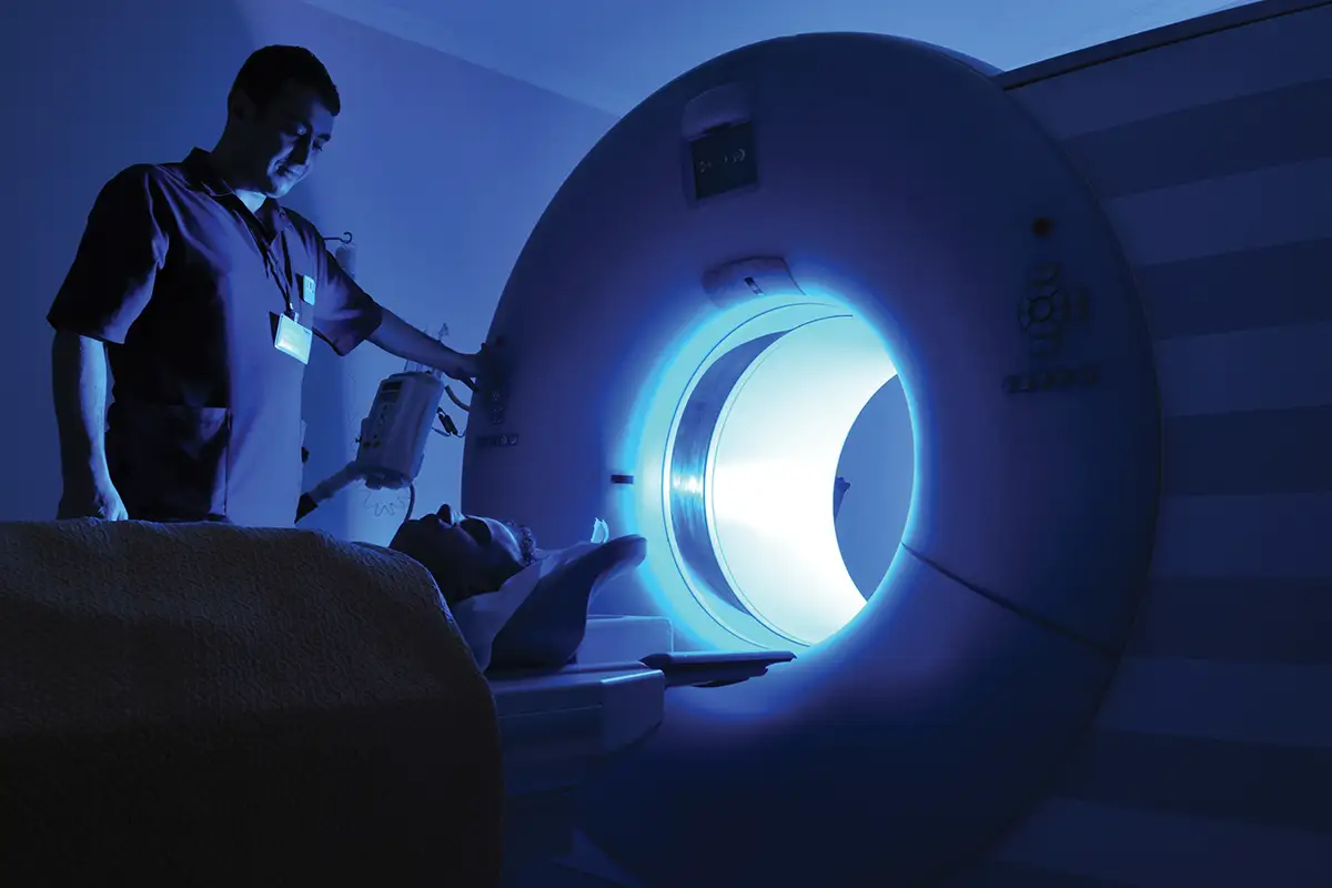
As MRI access expands around the world, the infrastructure needed to support safe, consistent scanning does not always grow at the same pace. That gap can become particularly consequential when a patient has an implant, a history of surgery, or incomplete medical records.
Before proceeding, the MRI team may need to identify a device, locate manufacturer documentation, determine its MRI status, and translate a list of technical conditions into a workable scanning protocol. Without centralized information and clearly defined processes, what should be a routine safety assessment can become a prolonged search through paper files, websites, PDFs, and institutional knowledge.
A new collaboration involving NordInsight and clinical researchers in Nigeria is examining whether structured digital workflow support can make that process more reliable in resource-limited environments. The broader research initiative, based at Bayero University Kano and the Federal University of Health Sciences, Azare, is supported by a 2026 Medical Image Computing and Computer Assisted Intervention Society Health Equity Grant and is exploring the development of AIassisted tools for MRI safety screening and protocol optimization.
NordInsight, a Copenhagen-based MRI safety technology company, has provided access to its platform to support implant evaluation and workflow standardization. The immediate goal, however, is not to test an autonomous AI system or validate a finished clinical product. It is to understand how MRI personnel work, where safety-related delays occur, and whether more structured access to information can reduce friction at the point of care.
“The collaboration was prompted by a practical safety and workflow problem,” says Simon Elliott Thomassen, cofounder and CEO of NordInsight. “MRI teams are increasingly seeing patients with implants, devices, prior surgery, or incomplete medical histories, but they do not always have fast access to reliable, device-specific MRI safety information at the point of care.”
A Fragmented Search Process
The challenge is familiar to MRI professionals in virtually every health care system. A screening form may indicate that a patient has an implanted device, but the patient may not know its manufacturer or model. Surgery may have been performed at another facility, and records may be unavailable or incomplete. Documentation may exist but be scattered among manufacturer manuals, online databases, local policies, and prior safety determinations. In settings with fewer personnel, less-developed IT systems, and limited access to dedicated MRI safety specialists, the burden of resolving those questions can fall on already stretched clinical teams.
“In Nigeria and similar settings, this can be especially difficult when implant documentation is incomplete, the patient does not know the exact device model, surgery took place at another facility, or staff need to piece together information from paper records, PDFs, web searches, manufacturer documents, and local experience,” Thomassen says.
The consequences can include delayed examinations, canceled appointments, inconsistent decisions, or unnecessary escalation of cases to radiologists, physicists, or other specialists. In some instances, a scan that could have been performed safely may be postponed because the team cannot confirm the required conditions efficiently.
Thomassen cautions against assuming that every Nigerian MRI department faces the same circumstances or attempting to quantify the problem without local data. Learning how the challenges appear within actual clinical workflows is one of the initiative’s purposes.
“This is not only a Nigerian issue,” he says. “Implant-related MRI safety screening is a global operational challenge. It becomes more difficult in settings where staffing, training resources, IT infrastructure, and access to centralized safety information are more limited.”
Learning Before Validating
Although early descriptions of the initiative emphasized AI-assisted screening and protocol optimization, the current work is best understood as an exploratory collaboration. NordInsight has made its platform available free of charge so the MRI team can determine where, and whether, it is useful in routine operations.
The initiative has not yet established predefined clinical scenarios, formal endpoints, or a structured validation protocol. No claims are being made regarding sensitivity, specificity, false-positive rates, or false-negative rates. Instead, the collaborators are looking at practical questions such as which cases generate the most difficult searches or what information is most often missing.
“The purpose right now is to understand how the product is used in a real MRI department: when staff choose to use it, what types of questions they use it for, where it fits naturally into their workflow, and where it does not,” Thomassen says.
The present phase centers on NordInsight and Bayero University Kano. The hospital team can use the platform when researching implants, reviewing MRI conditions, or determining whether a more organized information source can reduce the time spent consulting fragmented materials. More formal research, structured data collection, or clinical evaluation could be developed later with local partners and the necessary approvals.
That distinction is important in a field where the term “AI” can imply a level of automation that the project is not seeking to introduce. Thomassen says the long-term purpose is to support qualified personnel, not transfer MRI safety decisions to an algorithm.
“AI and structured data may eventually help with parts of the screening process that are currently manual, repetitive, or difficult to manage consistently,” he says. “That could include searching for implant information, interpreting incomplete device details, surfacing relevant MRI safety conditions, or helping staff see what information may still be missing.”
The final determination would remain with the institution and its clinical staff. “The important point is that the technology is intended to support MRI teams, not make autonomous safety decisions,” Thomassen says. “MRI safety decisions remain the responsibility of qualified clinical personnel and the hospital’s own safety process.”
From Device Status to Scan Planning
Identifying whether an implant is MR Safe, MR Conditional, or MR Unsafe is only one part of the process. For MR Conditional devices, the team must also determine whether the examination can be performed within the specified parameters. Those conditions may involve magnetic field strength, transmit coil selection, anatomical region, specific absorption rate, B1+rms limits, scan duration, patient positioning, or sequence restrictions. The operational challenge is converting technical documentation into a practical plan for a particular patient, scanner, and examination.
“We want to better understand the gap between ‘This implant is MR Conditional’ and ‘This is how the examination can be planned safely on this scanner under these conditions,’” Thomassen says.
For that reason, protocol optimization within the initiative does not mean allowing software to independently select or alter imaging sequences. It refers more broadly to helping staff locate and organize the conditions that must be considered during scan planning.
The current exploratory phase is not measuring protocol optimization as a formal endpoint. It is evaluating whether the platform can help personnel access the relevant information and apply it more consistently when preparing examinations for patients with implants or uncertain implant histories. The distinction reflects a central tension in MRI safety technology: A useful system must provide enough structure to reduce variability without obscuring the role of local policy, scanner configuration, institutional governance, and professional judgment.
“Standardization does not mean every hospital must make the same decision in every case,” Thomassen says. “Local policy, clinical judgment, scanner setup, available expertise, and institutional governance remain essential.”
What can potentially be standardized is the process leading to the decision: collecting the same core information, checking device details against available documentation, identifying applicable scanner and protocol restrictions, recording missing information, and documenting the rationale for proceeding, postponing, or escalating the case.
In the Nigerian collaboration, NordInsight is not imposing a predetermined workflow. The company is observing how the clinical team uses the platform and whether it can make the hospital’s existing processes more transparent and consistent. A later phase could examine documentation templates, escalation pathways, or more defined workflow models.
Designing for the Frontline
Any technology intended for a resource-limited environment must account for the fact that the department may not have a large implementation team, extensive training capacity, or an MRI safety expert available for every case. “The tool is being designed around the reality that many MRI departments do not have unlimited staff, dedicated MRI safety specialists available at all times, or large IT implementation teams,” Thomassen says. “It needs to be lightweight, easy to learn, and useful for frontline MRI staff.”
The platform’s design emphasizes structured workflows, clear presentation of available information, visibility of missing data, and straightforward documentation. The goal is to reduce the number of disconnected searches that staff must conduct rather than introduce another complex system requiring substantial technical support.
That philosophy may prove as relevant to high-resource health systems as it is to emerging MRI programs. Large organizations also struggle with fragmented device documentation, inconsistent screening procedures, multiple imaging sites, high patient volumes, and differing levels of staff experience. In those systems, the problem is often not the absence of expertise but the difficulty of making that expertise available consistently across locations and shifts.
“A tool that works in a resource-limited setting has to be practical, simple, and efficient,” Thomassen says. “Those same qualities are valuable everywhere.”
Defining the Next Step
Success currently will be determined by what the collaborators learn rather than by a clinical performance statistic. The team will consider how often personnel use the platform, the kinds of cases for which they use it, whether it makes device information easier to locate, and where it fails to match the local workflow.
The collaborators are equally interested in identifying where the technology is not useful enough. That feedback could shape future product design and determine whether a structured pilot is warranted.
If the exploratory phase demonstrates clear value, a subsequent project could define specific use cases and evaluation criteria. It might focus on patients with known or suspected implants, map the hospital’s current safety process, provide staff training, collect structured feedback, and measure where the system produces the greatest operational benefit.
Broader implementation would require more than technical functionality. Data governance, affordability, regulatory requirements, training, institutional policies, and user trust would all influence whether the platform could be deployed across additional hospitals, regions, or countries.
“The long-term vision is not an autonomous MRI safety decision-maker,” Thomassen says. “It is a practical decision support and workflow infrastructure layer that helps MRI teams find the right information, understand what is missing, and reach clearer documented decisions without unnecessary delay.”
For MRI departments working with limited staff and scattered information, that means infrastructure may be as important as the intelligence built on top of it.
Keith Loria is a freelance writer based in Oakton, Virginia. He is a frequent contributor to Radiology Today.