


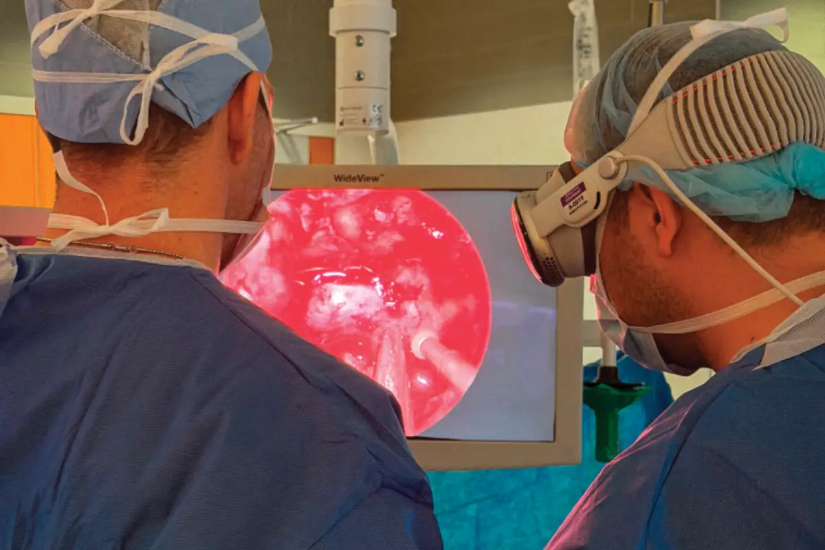
For decades, medicine has depended on a familiar process: acquire images, interpret them on a screen, mentally reconstruct anatomy in three dimensions, and translate that understanding into clinical decisions. Today, that model is evolving. Advances in AI, mixed reality, extended reality (XR), spatial computing, image fusion, and 3D disease mapping are transforming how clinicians interact with information, bringing complex anatomy, pathology, and procedural data directly into the clinical workspace.
Perhaps most remarkable is that this transformation is occurring simultaneously across multiple specialties. At the University of Pittsburgh, surgeons are exploring mixed reality systems that create immersive digital operating rooms. At Cleveland Clinic, interventional radiologists are using XR and image fusion to improve procedural navigation. In urologic oncology, AI-driven platforms such as Unfold AI are generating patient-specific cancer maps that support precision treatment planning and robotic surgery. Although these technologies differ in form and function, they share a common goal: transforming medical data into actionable clinical intelligence that enables more precise, personalized, and informed decision-making.
Translating Images to Anatomy
One of medicine’s longstanding challenges is translating 2D imaging into a meaningful 3D understanding of anatomy and disease. Advanced visualization technologies address this gap by integrating imaging, pathology, and procedural data into interactive, patient-specific models that clinicians can explore in real time. This goes beyond simply providing more information. These tools transform complex data into intuitive, clinically relevant visualizations that align with how clinicians naturally see, think, and make decisions, which is ultimately what drives their impact and value.
According to Adam Rakestraw, JD, cofounder, chairman, and chief business development officer for MediView XR, Inc, conventional imaging workflows often create a disconnect between clinicians, patients, and the anatomy being treated. The challenge is not a lack of data but how that data is delivered during critical moments of decision-making. “This isn’t a better picture,” he explains. “It’s the right picture, in the right place, at the right moment.”
This philosophy also guides the work of Edward Andrews, MD; Garret Choby, MD; and Georgios Zenonos, MD, at the University of Pittsburgh School of Medicine. Their mixed reality platform integrates imaging studies, surgical navigation, operative video, and physiologic data into a shared virtual workspace, allowing surgeons to access critical information without shifting their attention between multiple monitors. Choby notes, “In the integrated mixed reality platform, you have all those things in your view simultaneously, and you can also manipulate them.” Zenonos emphasizes that the platform’s value extends beyond visualization by giving surgeons direct control over imaging, navigation, and other digital resources within a single customizable environment.
A similar transformation is underway in cancer care. At Urology Cancer Specialists in Los Angeles, founder and medical director S. Adam Ramin, MD, uses Avenda Health’s Unfold AI to integrate MRI scans, biopsy results, and pathology findings into a patient-specific 3D model of prostate cancer. The resulting visualization helps clinicians better understand tumor location, extent, and potential spread beyond the prostate. Ramin says physicians need to know not only where a tumor is located but also whether it may extend beyond the prostate capsule. By combining multiple data sources, he says, “Unfold AI allows us to create a 3D map of the prostate cancer.”
Although these technologies serve different specialties, they share a common objective: giving clinicians a clearer and more intuitive understanding of anatomy and disease. By transforming complex datasets into interactive visual models, they help convert information into actionable clinical insight at the point of care.
Better Visualization
Perhaps the most immediate benefit of advanced visualization technologies is improved procedural precision. By providing a clearer understanding of anatomy, pathology, and instrument positioning, these platforms are helping reduce uncertainty during complex interventions.
In IR, the MediView XR90 Holographic Surgical Navigation System combines CT-derived anatomy with real-time ultrasound into a unified 3D roadmap. Rather than mentally merging separate imaging sources, clinicians can visualize both simultaneously. As Kevin Stadtlander, MD, interventional radiologist at Cleveland Clinic, explains, it is “a way to use two imaging modalities at once.”
By combining the anatomical detail of CT with the real-time feedback of ultrasound, the platform may improve targeting accuracy and procedural confidence. “The concept is to be more accurate in terms of guiding a needle exactly where you want it to go,” Stadtlander says. According to Rakestraw, the technology has already been used in more than 500 procedures across over 20 anatomical locations.
Precision is equally important in prostate cancer care. Using Unfold AI, clinicians can combine MRI findings, biopsy results, and pathology data into a patientspecific 3D cancer map that helps define disease extent and guide treatment planning. Because focal therapy targets only the cancerous portion of the prostate while preserving healthy tissue, accurate visualization is essential. “If we have a 3D image of the tumor, then we know which part of the prostate we can go and treat focally,” Ramin says. The technology also helps identify areas where treatment may need to extend beyond the prostate because of potential extracapsular disease.
In the operating room, mixed reality platforms developed by Andrews, Choby, and Zenonos, at the University of Pittsburgh integrate navigation systems, endoscopic imaging, and physiologic data into a shared virtual workspace. According to Choby, the technology changes how surgeons view information rather than how they perform surgery. “We can expand things, make them larger, smaller, move them here, move them there in our view,” he says. By placing critical information directly within the surgeon’s field of view, the platform reduces reliance on multiple monitors while supporting more informed intraoperative decision-making.
Improving Patient Outcomes
Ultimately, the value of advanced visualization technologies lies in their potential to improve patient outcomes through more precise and personalized care. In prostate cancer care, AI-generated 3D cancer maps help clinicians better understand tumor location and potential extracapsular extension, supporting more targeted treatment while preserving healthy tissue. This is especially important during nerve-sparing robotic prostatectomy, where surgeons must balance cancer removal with preservation of urinary and sexual function.
“It can potentially help us as surgeons be able to get negative margins, be able to resect the tumor better, and at the same time give our patients a better quality of life in terms of their sexual function and bladder function,” Ramin says.
In IR, platforms such as the MediView XR90 combine CT-derived anatomy with real-time ultrasound to improve target localization and procedural accuracy. Greater precision may reduce needle passes, shorten procedure times, and lower complication rates while reducing reliance on repeated CT confirmation scans. Stadtlander believes this approach may be an important step toward reducing dependence on radiation-intensive guidance methods and could eventually expand to procedures that currently rely on fluoroscopy.
Benefits extend into the operating room, as well. At the University of Pittsburgh, researchers envision mixed reality environments that place navigation data, imaging studies, physiologic information, and future AI-driven insights directly within the surgeon’s field of view. By improving access to critical information during procedures, these systems may support faster decision-making, greater precision, and better preservation of surrounding anatomy and function.
The potential of this technology is perhaps best illustrated through the experience of its first patient. Amanda McGrail, a 38-year-old mechanical and aerospace engineer from Maryland, underwent endoscopic endonasal surgery at the University of Pittsburgh Medical Center for a pituitary tumor causing Cushing’s disease. After nearly a year of seeking answers for symptoms that included chronic anxiety, profound fatigue, unexplained weight gain, and a significant decline in daily functioning, McGrail became the first patient to undergo surgery using the mixed reality platform developed by Andrews, Choby, and Zenonos. Although she understood she was participating in a first-of-itskind procedure, McGrail was enthusiastic about the opportunity.
“Dr. Andrews called me the night before surgery and explained what they were working on,” she says. “As an engineer, I was fascinated. My only question was whether there was still a backup plan, if needed. Once I knew there was, I was all in. Someone has to go first.”
Following surgery, McGrail experienced a dramatic improvement in her symptoms. She later realized the extent to which Cushing’s disease had affected her daily life when she was able to navigate a stressful insurance issue without the anxiety that had previously become routine. For her, recovery was better than expected, and she now encourages other patients facing similar diagnoses to consider surgery when recommended by their physicians.
Although further research is needed to quantify their impact, the promise of these technologies is clear: helping clinicians make better decisions and deliver more precise, patient-centered care. Whether through reducing radiation exposure, preserving quality of life after cancer treatment, minimizing complications, or improving procedural accuracy, advanced visualization technologies are becoming tools not just for seeing anatomy more clearly but also for improving outcomes.
AI and Clinical Decision-Making
Yet visualization is only part of the story. As these platforms evolve, AI is increasingly being used to organize, interpret, and contextualize complex clinical data, transforming visualization systems into decision-support tools that help clinicians make faster, more informed decisions.
In prostate cancer care, AI is already influencing treatment planning. Platforms such as Unfold AI analyze MRI findings, biopsy results, and pathology data to generate predictive 3D maps that help clinicians assess disease extent and personalize treatment strategies across focal therapy, radiation oncology, active surveillance, and surgery. For Ramin, the value of AI lies in its ability to transform disparate clinical data into an actionable representation of disease, eliminating the need to mentally integrate multiple data sources during decision-making.
At the University of Pittsburgh, researchers envision AI evolving far beyond image interpretation. Their goal is an intelligent operating room where AI functions as a real-time clinical assistant. In Andrews’ vision, AI could continuously analyze operative data and provide relevant information when it is needed, helping support decision-making without increasing cognitive burden. The concept of a “surgical copilot” would allow clinicians to access critical insights without actively searching for them.
A similar future is emerging in procedural medicine. According to Rakestraw, platforms such as MediView’s XR90 system generate far more than visual guidance, capturing 3D spatial procedural data related to anatomy, instrument location, motion, workflow, and imaging relationships. This information will provide the foundation for advanced AI applications within MediView’s Clinical Spatial Intelligence ecosystem.
“Every tracked procedure produces an unprecedented dataset,” Rakestraw says. “That’s the foundation for the next era of procedural intelligence that links imaging, planning, navigation, and therapy-specific applications.” Together, these technologies are laying the groundwork for AI-enabled clinical environments that can learn from procedures, improve workflows, and provide increasingly personalized support.
Reducing Cognitive Burden
At the University of Pittsburgh, researchers found that surgeons could access multiple data streams simultaneously without increasing mental burden. “The system doesn’t seem to offer any increased cognitive load for the surgeon,” Andrews says.
A similar experience has been reported in IR. Despite integrating CT imaging, real-time ultrasound, image fusion, and extended reality visualization, the Medi- View XR90 platform fits into existing workflows. “Workflow is exactly the same as doing the procedure, just with a handheld ultrasound,” Stadtlander says.
By presenting relevant information within a unified environment, advanced visualization platforms reduce the effort required to interpret and integrate multiple data sources. Instead of shifting attention between monitors, imaging systems, and patient data, clinicians can spend more time focused on patient care and procedural decisions.
Many clinicians also report ergonomic benefits. By placing imaging, navigation data, and procedural guidance directly within the user’s field of view, mixed and XR systems reduce the need to repeatedly turn toward external displays. For Choby, one of the most immediate advantages is flexibility in the operating room. “When the goggles are on your face, you can stand however you want to. The surgery is in your field of view,” he says.
Similarly, Stadtlander notes, “You’re able to look at the patient. You’re seeing through your headset what you need to see, and you don’t need to look away at another monitor.” According to Rakestraw, internal evaluations of the MediView XR90 demonstrated approximately 47% less ergonomic strain than conventional workflows.
From Static Images to Interactive Intelligence
Although the clinical applications differ, the experts interviewed described remarkably similar visions for the future. They foresee technologies that are increasingly immersive, intelligent, predictive, and interconnected.
Andrews imagines digital operating rooms where AI and spatial computing support surgical decision-making in real time. Choby views today’s mixed reality platforms as an important foundation for a future in which physical monitors may no longer be necessary. “It’s the first step in the future of how you view a surgical field in virtual reality,” he says.
Zenonos views mixed reality as more than a replacement for traditional monitors. He sees it as a flexible digital platform capable of continuously incorporating future innovations, from advanced imaging and navigation technologies to AI-driven decision support.
Stadtlander foresees the emergence of “augmented interventional radiology,” where XR, AI, robotics, digital twins, and advanced procedural simulation converge to expand physician capabilities. In his view, the future lies not in any single technology, but in the integration of multiple technologies that provide increasingly sophisticated visual, navigational, and analytical support.
Ramin hopes future systems move beyond side-by-side displays and allow AI-generated models to be projected directly onto surgical anatomy through augmented reality overlays. Looking ahead, he envisions a time when surgeons can view AI-generated cancer maps directly on the operative field itself, creating a more intuitive connection between digital information and patient anatomy.
“I would also like to be able to have more of an augmented reality-type image where we can project the AI image directly onto the prostate video,” he says.
What connects these diverse innovations is their shared movement away from passive visualization and toward interactive clinical intelligence. Instead of requiring clinicians to search for information, future systems may deliver information contextually, displaying anatomy, pathology, procedural guidance, and predictive insights precisely when they are needed.
The next era of health care may no longer be defined by what clinicians can see on a screen. Instead, it may be defined by how effectively technology helps them understand disease, navigate anatomy, make decisions, and deliver truly personalized care. The future of visualization is not simply about seeing more. It is about transforming information into understanding, understanding into action, and action into better outcomes for patients.
Rebecca Montz, EdD, MBA, CNMT, PET, RT(N)(CT), NMTCB RS, has worked at the Mayo Clinic Jacksonville and University of Texas MD Anderson Cancer Center in Houston as a nuclear medicine and PET technologist.