


History
A 27-year-old man with a history of asthma, recurrent nephrolithiasis, and urethral stricture treated with dilation presented to the emergency department with three days of left lower quadrant and rectal pain, described as pressure and tearing with bowel movements, and one to two weeks of dysuria with red-tinged urine. He also reported chills and nausea. A pelvic mass identified on CT at an outside hospital two months earlier had increased in size on repeat outside CT the day before presentation. On arrival, he was afebrile with a normal white blood cell count, and serum carcinoembryonic antigen and alpha-fetoprotein levels were normal. Urinalysis was mildly abnormal, and he was treated for presumed cystitis. Contrast- enhanced CT of the abdomen and pelvis was obtained, and he was admitted for pain control. Pelvic MRI followed.
Findings
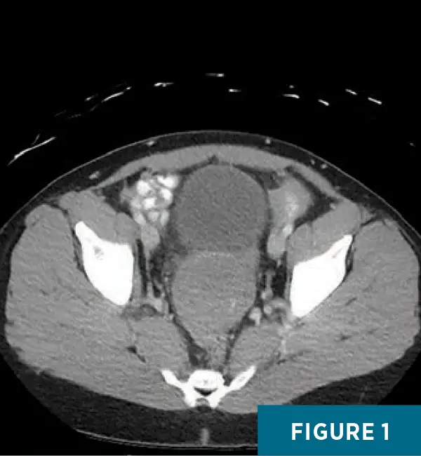
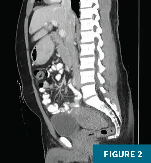
Contrast-enhanced CT of the abdomen and pelvis demonstrated an ill-defined, mildly heterogeneous soft-tissue mass measuring 6.8 X 6.3 X 5.3 cm (anteroposterior X transverse X craniocaudal), appearing to arise from the anterior aspect of the sigmoid colon (Figures 1 and 2). The mass demonstrated central hypoattenuation with peripheral enhancing solid components, suggestive of necrosis or cystic change within a viable tumor rim. There was no evidence of bowel obstruction.
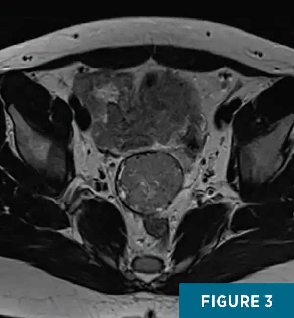
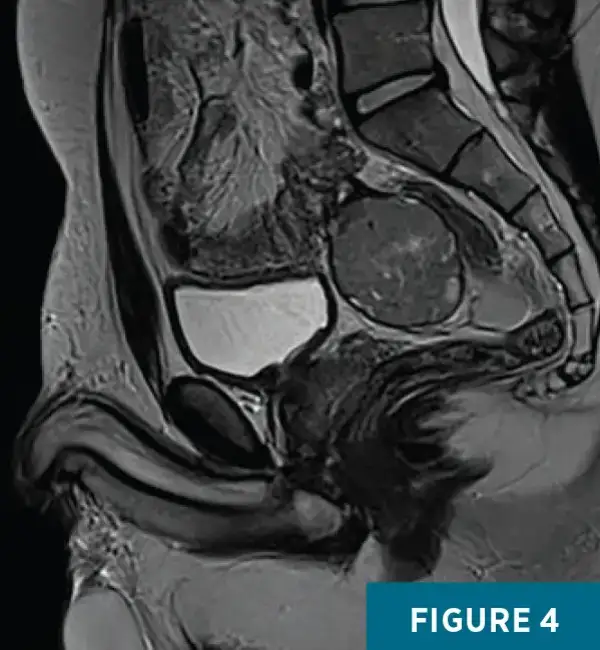
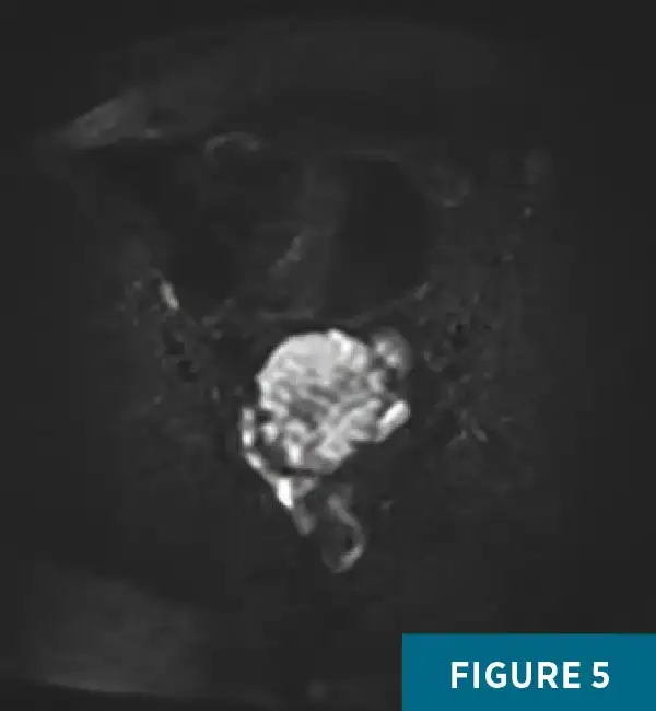
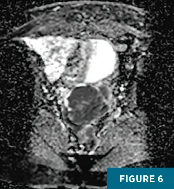
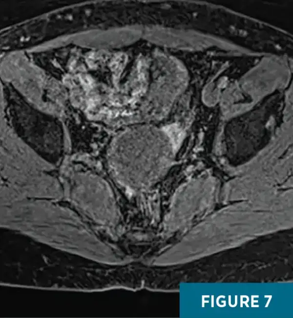
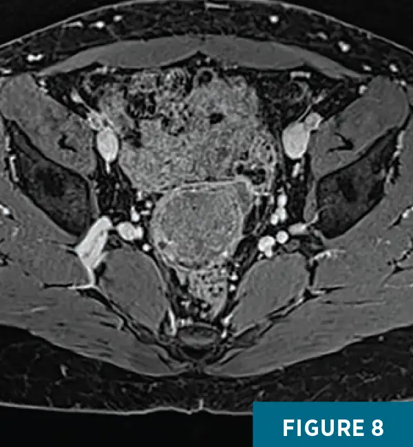
Noncontrast chest CT demonstrated no evidence of metastatic disease. Pelvic MRI with and without contrast redemonstrated the heterogeneous mass arising from the distal sigmoid colon, posterior to the bladder. The lesion demonstrated heterogeneous T2-hypointense signal (Figures 3 and 4), areas of restricted diffusion (Figures 5 and 6), and mild enhancement following contrast administration (Figures 7 and 8).
There was no significant signal dropout on opposed-phase imaging. The mass was separate from the seminal vesicles and prostate. A small amount of nonenhancing, T1-hyperintense fluid surrounded the mass, likely representing proteinaceous or hemorrhagic material. The remaining visualized bowel, bladder, and prostate were unremarkable, without bowel obstruction or pelvic lymphadenopathy.
This constellation of imaging findings suggested that the sigmoid colon was the tissue of origin, with features most consistent with a gastrointestinal mesenchymal neoplasm, of which gastrointestinal stromal tumor (GIST) is the most common. Colonic adenocarcinoma was considered less likely, as the mass did not appear intraluminal and there was no appreciable lymphadenopathy.
Following MRI, sigmoidoscopy with biopsy and immunohistochemical analysis revealed a KIT (CD117)–positive GIST. The patient continued to experience abdominal discomfort, particularly with bowel movements, but denied hematochezia.
Diagnostic laparoscopy was subsequently performed to assess resectability. Blood staining was present throughout the peritoneal cavity, with fluid concentrated around the mass, which was adherent to the rectum and a loop of small bowel and appeared fused to the bladder—findings concerning for tumor perforation.
Resection was deferred, as it was deemed more appropriate to initiate systemic therapy for tumor reduction before proceeding with low anterior resection. Following laparoscopy, the patient was started on imatinib and remains under surveillance by colorectal surgery and oncology. Given his young age at onset, genetic evaluation is planned to assess for a hereditary predisposition.
Diagnosis
GIST of the sigmoid colon
Discussion
Although uncommon, GISTs are the most frequent mesenchymal neoplasms of the gastrointestinal tract. They are typically composed of spindle cells and, less commonly, epithelioid cells.1 The annual incidence is approximately 11 to 15 cases per million. About 55% of GISTs arise in the stomach and 31% in the small intestine, whereas only about 6% occur in the colon or rectum2; colorectal GISTs, including those in the sigmoid colon, are therefore rare.3 Incidence peaks in the seventh decade of life,2 with no clear sex predilection.
Approximately 75% to 80% of GISTs harbor activating KIT mutations, 5% to 10% harbor platelet-derived growth factor alpha mutations, and more than 95% express KIT (CD117), reflecting their origin from the interstitial cells of Cajal, the pacemaker cells of the gastrointestinal tract.1,2 Although most GISTs are sporadic, germline KIT mutations (familial GISTs) may result in multiple tumors or diffuse hyperplasia of Cajal cells.1 Other hereditary associations include succinate dehydrogenase-deficient GISTs (seen in Carney–Stratakis syndrome) and neurofibromatosis type 1.2 Presentation at a young age without other risk factors should raise suspicion for an underlying hereditary syndrome and prompt genetic evaluation.
Small GISTs (<2 cm) are often asymptomatic; larger lesions may present with abdominal pain, bleeding, or obstruction. In colorectal locations, symptoms may include altered bowel habits, pelvic discomfort, or urinary symptoms from mass effect, as in this case.3
The differential diagnosis for a pelvic or colonic mass includes:
• Adenocarcinoma: an irregular, infiltrative mass causing circumferential wall thickening and luminal narrowing, often with pericolonic fat stranding and lymphadenopathy.
• Neuroendocrine tumor: a well-defined, hypervascular submucosal mass, sometimes with mesenteric desmoplastic reaction; best evaluated with somatostatin receptor PET/CT (eg, gallium Ga 68 dotatate).
• Lymphoma: a homogeneous bulky mass or diffuse mural thickening with frequent nodal involvement but less obstruction.
• Desmoid tumor: a soft-tissue mass that may appear well circumscribed but is locally infiltrative, often associated with familial adenomatous polyposis or prior surgery.
• Leiomyoma/leiomyosarcoma: leiomyomas are small, well-circumscribed intramural masses with low attenuation on CT and low T2 signal on MRI; leiomyosarcomas are larger and more heterogeneous and are far less common than GISTs.
• Metastases: variable in appearance depending on primary malignancy, requiring correlation with clinical history.
Contrast-enhanced CT is the primary modality for diagnosis and staging, and MRI is indicated for rectal GISTs.2 GISTs typically appear as well-defined endoluminal or exophytic masses with heterogeneous enhancement due to necrosis. Aggressive features include large size (>10 cm), irregular margins, necrosis, and invasion of adjacent structures.4 MRI shows hypo- to isointense T1 and heterogeneous T2 signal, reflecting necrosis or hemorrhage. PET/CT is used selectively, chiefly to assess early response to imatinib.2
Histopathologic confirmation with KIT (CD117) immunopositivity establishes the diagnosis, as normal colonic smooth muscle is KIT-negative; DOG1 staining identifies about half of KIT-negative GISTs. Recurrence risk is stratified by tumor size, mitotic count (>5 per 50 high-power fields), anatomic site, and tumor rupture.2
The cornerstone of management is complete surgical resection with negative margins (R0), which offers the best prognosis. For localized sigmoid GISTs, segmental colectomy with mesenteric resection is typically performed; lymphadenectomy is usually unnecessary due to the rarity of nodal spread.2,3 Tyrosine kinase inhibitors such as imatinib mesylate are indicated as adjuvant therapy for high-risk disease and as first-line treatment for unresectable or metastatic tumors. Neoadjuvant imatinib may facilitate resection in large or borderline operable lesions.2,5
After complete resection, approximately 40% of patients develop metastatic relapse, most commonly to the liver and peritoneum.2 Relapse occurs most often within two years after adjuvant imatinib is discontinued. Five-year relative survival is approximately 96% for localized disease, 87% for regional spread, and 57% for distant metastases.6 Surveillance CT is recommended every four to six months for at least five years in intermediate- and high-risk cases.2
Sigmoid GISTs are rare tumors arising from the interstitial cells of Cajal that typically present with nonspecific symptoms or are discovered incidentally on imaging. Diagnosis relies on characteristic imaging findings and histopathologic confirmation of KIT positivity. Surgical excision remains the mainstay of therapy, complemented by imatinib for high-risk or metastatic disease. Despite advances, recurrence remains common, emphasizing the need for long-term imaging surveillance.
Youssef Beshay, MD, is a radiology resident at UConn Health in Farmington, Connecticut.
Eyad Nagori is a medical student at the University of Connecticut School of Medicine in Farmington, Connecticut.
Alex Merkulov, MD, is an associate professor of radiology at UConn Health.
References
1. Kumar V, Abbas AK, Aster JC, eds. Robbins and Cotran Pathologic Basis of Disease. 9th ed. Elsevier; 2015:775-777.
2. Serrano C, Martín-Broto J, Asencio-Pascual JM, et al. 2023 GEIS guidelines for gastrointestinal stromal tumors. Ther Adv Med Oncol. 2023;15:17588359231192388.
3. Reddy RM, Fleshman JW. Colorectal gastrointestinal stromal tumors: a brief review. Clin Colon Rectal Surg. 2006;19(2):69-77.
4. King DM. The radiology of gastrointestinal stromal tumours (GIST). Cancer Imaging. 2005;5(1):150-156.
5. Maki RG. Gastrointestinal stromal tumors (GIST) and their management. Gastrointest Cancer Res. 2007;1(4 suppl 2):S81-S84.
6. Survival rates for gastrointestinal stromal tumors. American Cancer Society website. https://www.cancer.org/cancer/types/gastrointestinal-stromal-tumor/detection-diagnosis-staging/survival-rates.html. Updated March 10, 2026. Accessed October 20, 2026.