


A 57-year-old woman presented for evaluation of a suspected right breast abscess at the 10 o’clock position. Her primary care provider had noted a raised, inflamed right breast mass on physical examination. The patient had completed a seven-day course of oral antibiotics without improvement, and the lesion had been present for approximately two months. Bilateral mammography and targeted ultrasound of the right breast were obtained.
Her medical history was notable for a long-standing overlap connective tissue disease—predominantly dermatomyositis with features of systemic lupus erythematosus and scleroderma—diagnosed more than 20 years earlier and complicated by calcinosis cutis. A high-resolution chest CT obtained in 2023 demonstrated diffuse heterogeneous subcutaneous calcifications throughout the chest wall, without interstitial lung disease. Her connective tissue disease had been managed with long-term immunosuppressive therapy, including corticosteroids and intravenous immunoglobulin. Additional history included hypothyroidism, diabetes mellitus, and osteopenia.
Findings
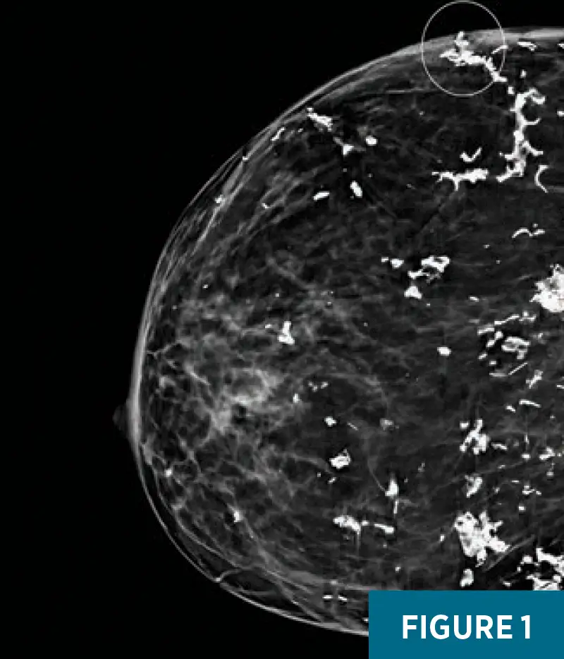
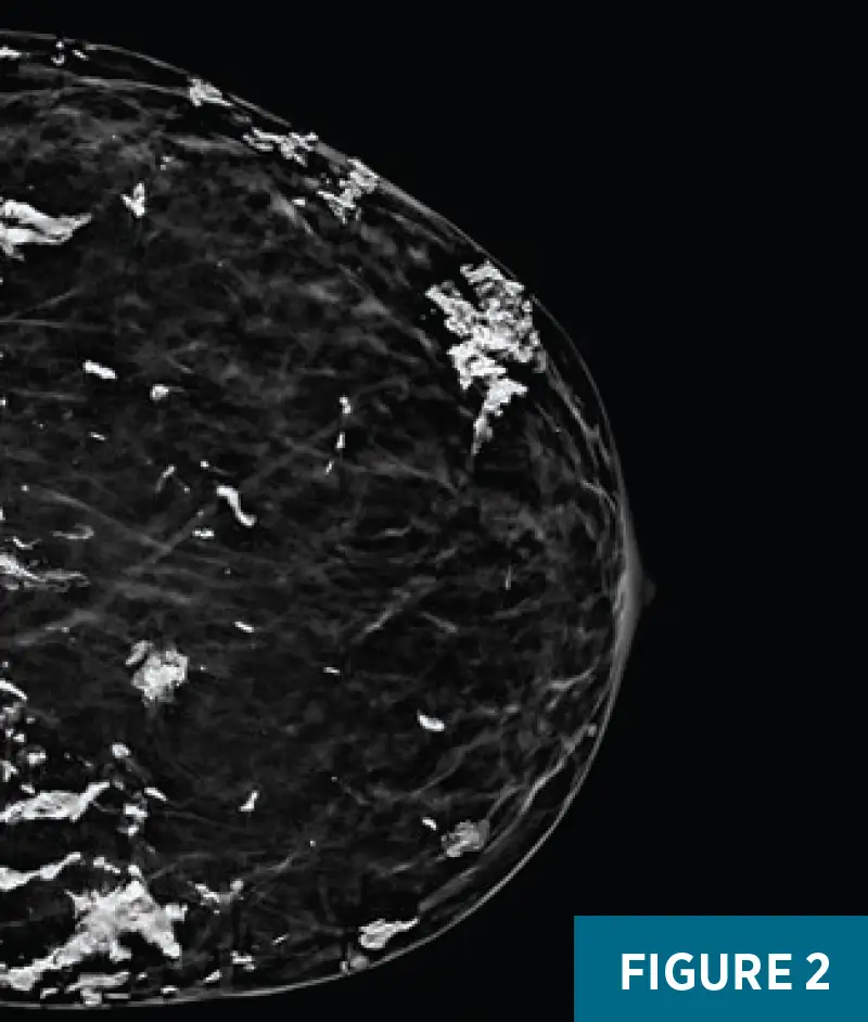
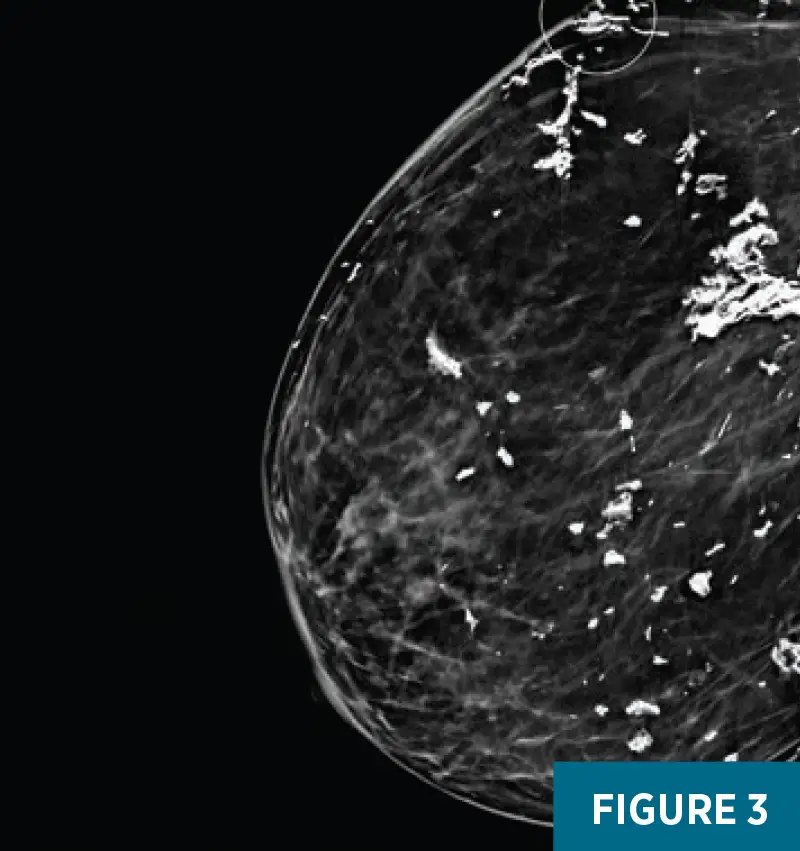
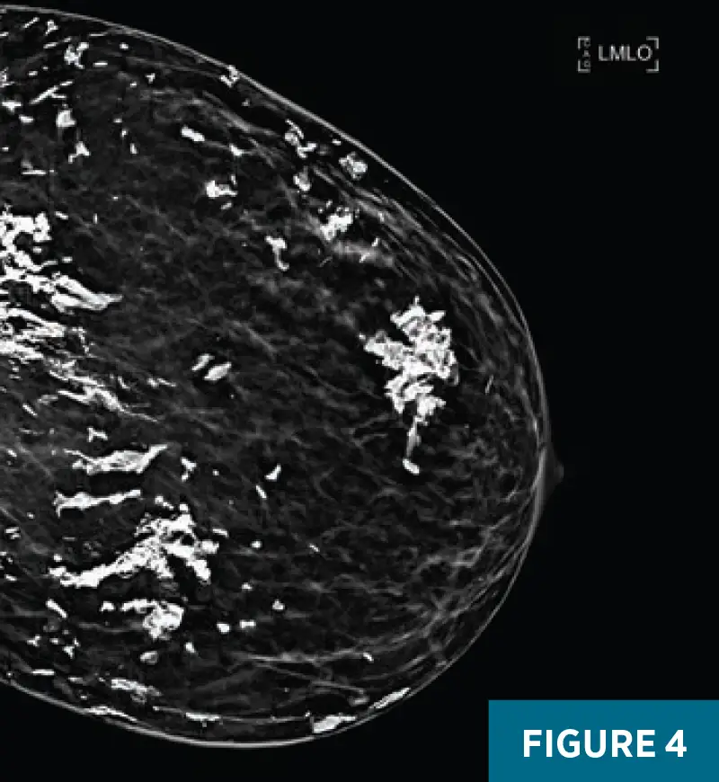
Figures 1–4. Bilateral craniocaudal and mediolateral oblique mammographic views demonstrate diffuse, coarse, morphologically benign dermal and subcutaneous calcifications. On the right breast craniocaudal and mediolateral oblique views (Figures 1 and 3), a coarse, subcutaneous calcification is present at the 10 o’clock position, 13 cm from the nipple, extending through the dermis, with mild focal dermal thickening.
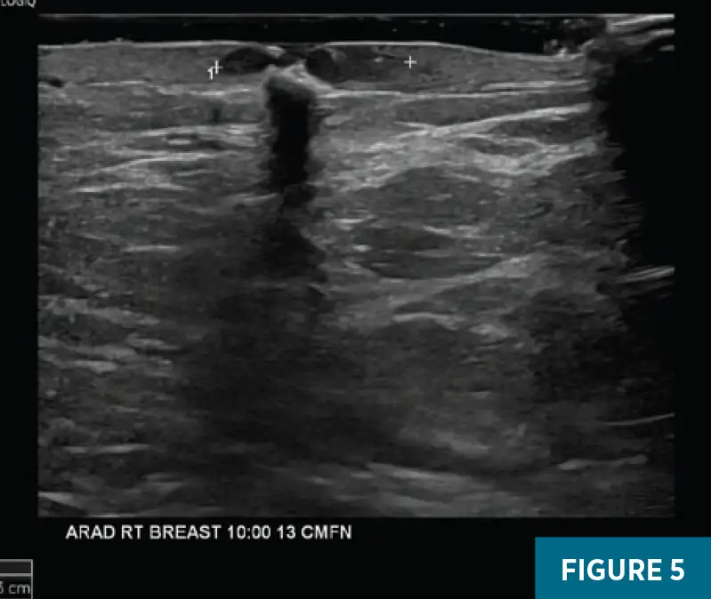
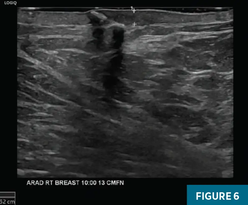
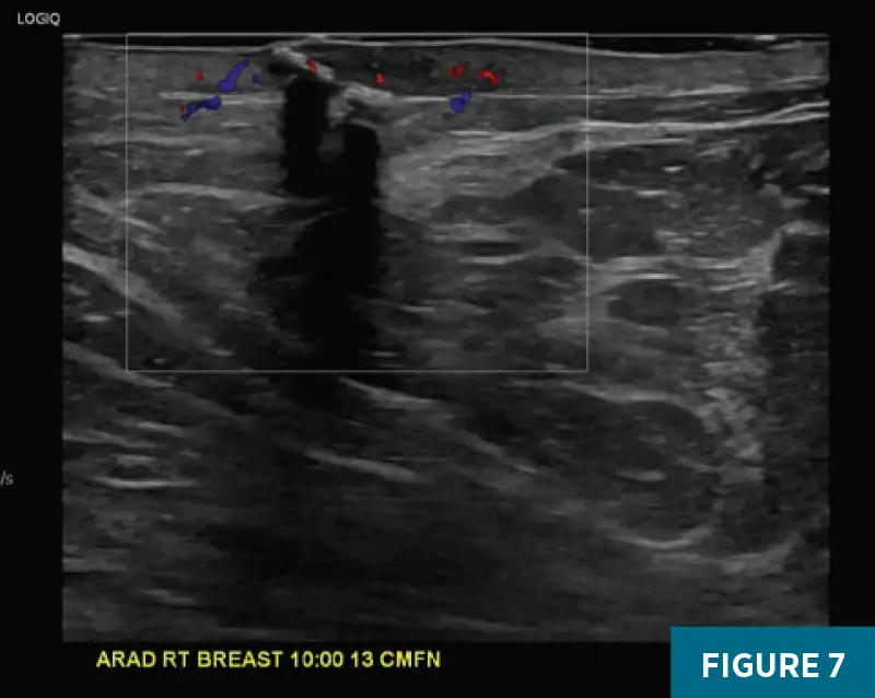
Figures 5–7. Targeted ultrasound of the right breast demonstrates a coarse, subcutaneous calcification breaching the dermis, associated with a 1.5 cm hypoechoic focus of dermal inflammation and mild dermal thickening measuring up to 3 mm; no subcutaneous abscess is identified.
Based on the imaging findings and clinical presentation, the diagnosis of infected or sterile calcinosis and a focal foreign-body granulomatous reaction was established, and a multidisciplinary management approach was recommended. At follow-up with internal medicine approximately three months later, the right breast dermal lesion had resolved completely without further intervention. Topical sodium thiosulfate cream had been prescribed for the calcinosis, but the patient was unable to obtain it because of insurance limitations.
Diagnosis
Breast calcinosis in a patient with dermatomyositis
Discussion
Dermal lesions are commonly encountered in the breast imaging clinic and represent a wide spectrum of conditions. They are often incidental findings on mammography or ultrasound and may mimic underlying parenchymal pathology if not correctly identified. Common dermal entities include epidermal inclusion cysts, sebaceous cysts, dermal calcifications, and skin tags.
The differential diagnosis for a raised, inflamed skin lesion of the breast, as seen in our patient, is broad and includes infectious, inflammatory, autoimmune, and neoplastic causes. Common benign etiologies include mastitis, breast abscess, and infected epidermal inclusion cysts, typically presenting with localized pain, erythema, and tenderness. Granulomatous mastitis and hidradenitis suppurativa may cause chronic, recurrent, or sinus-forming lesions. Autoimmune and connective tissue diseases such as scleroderma and dermatomyositis can present with calcinosis cutis, which may ulcerate. Importantly, inflammatory breast carcinoma and cutaneous metastases should be considered nonresolving or rapidly progressive lesions that mimic infection but fail to respond to antibiotics.
Given our patient’s history of connective tissue disease, breast calcinosis was considered the most likely cause of the diffuse, coarse calcifications on her imaging. Breast calcinosis, or more broadly calcinosis cutis, refers to the abnormal deposition of calcium salts, most often calcium phosphate, in the skin and soft tissues. It can manifest in various forms, ranging from small, firm subcutaneous nodules to larger, more extensive deposits, and is a well-recognized but variable manifestation of systemic connective tissue disease. It occurs most frequently in limited systemic sclerosis and juvenile dermatomyositis, occasionally in mixed connective tissue disease and adult dermatomyositis, and is rare in systemic lupus erythematosus or rheumatoid arthritis. It usually reflects chronic inflammation, vascular damage, or tissue necrosis. As a sequela of connective tissue disease, calcinosis is often associated with early-onset disease and suboptimal or delayed treatment. Deposited calcium may ulcerate and discharge chalky material, often complicated by secondary infection. In systemic connective tissue disease, calcinosis cutis most commonly develops in areas subject to repeated trauma or pressure, particularly over joints and bony prominences; typical sites include the fingers, elbows, forearms, knees, and buttocks. Breast involvement is relatively rare.
On mammography, breast calcinosis typically presents as coarse, dystrophic dermal and subcutaneous calcifications. Ultrasound may reveal echogenic foci with posterior acoustic shadowing within the dermis or subcutaneous tissues, corresponding to calcified deposits, and dermal thickening may also be noted. CT and plain radiography can delineate the extent and density of calcium deposition, while MRI is less sensitive for detecting calcification but may help assess associated inflammation or soft-tissue involvement. Diagnosis is usually based on clinical evaluation, imaging, and pathological confirmation demonstrating calcium deposits in the dermis or subcutaneous tissues without underlying malignancy. Management is often challenging and centers on treating the underlying systemic disease, alongside pain control and prevention of infection. Medical therapies may include diltiazem, minocycline, bisphosphonates, or sodium thiosulfate (topical, intralesional, or intravenous), though responses are variable. In cases of ulceration, local wound care and infection control are essential, and surgical excision or debridement may be considered for symptomatic or refractory lesions.
In summary, a wide variety of dermal lesions are evaluated by breast radiologists. Calcinosis cutis is a well-known cutaneous manifestation of systemic connective tissue disease but is rare in the breast. A combination of careful clinical evaluation, imaging, and histopathology is recommended for accurate diagnosis. Management is multidisciplinary, focusing on controlling the underlying disease, symptom relief, infection prevention, and medical or surgical intervention for ulcerated or refractory lesions.
— Arnika Karthik, MD, is a radiology resident at UConn Health in Farmington, Connecticut.
— Alex Merkulov, MD, is an associate professor of radiology at UConn Health.
Resources
1. Balin SJ, Wetter DA, Andersen LK, Davis MDP. Calcinosis cutis occurring in association with autoimmune connective tissue disease: the Mayo Clinic experience with 78 patients, 1996-2009. Arch Dermatol. 2012;148(4):455-462.
2. Gutierrez A Jr, Wetter DA. Calcinosis cutis in autoimmune connective tissue diseases. Dermatol Ther. 2012;25(2):195-206.
3. Namas R, Beydoun N, Meysami A. Breast calcinosis in a patient with dermatomyositis. Eur J Rheumatol. 2017;4(2):175-176.
4. Sewchuran T, Kabeya JM. Calcinosis circumscripta of the breasts: the deeper meaning. SA J Radiol. 2023;27(1):2730.
5. Bin Saeedan M, Mobara M, Arafah MA, Mohammed TL. Breast lesions on chest computed tomography: pictorial review with mammography and ultrasound correlation. Curr Probl Diagn Radiol. 2015;44(2):144-154.