


History
A 34-year-old nulliparous woman presented with a self-detected right breast lump noticed approximately six weeks earlier. The lump had remained unchanged and was not painful, with intermittent itching and mild darkening of the overlying skin but no erythema, drainage, or fever. Her history was notable for obesity, with no prior breast disease, ipsilateral skin infection, or family history of breast or ovarian cancer. On referral for surgical evaluation, examination demonstrated a firm, slightly immobile right breast mass at the 10 o’clock position near the mid axillary line, without fluctuance or erythema and with no findings supporting infection. The patient was then referred for a diagnostic mammography and ultrasound and subsequently underwent an ultrasound-guided breast mass biopsy.
Findings
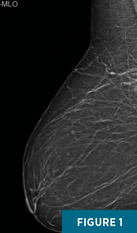
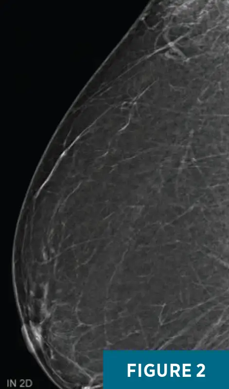
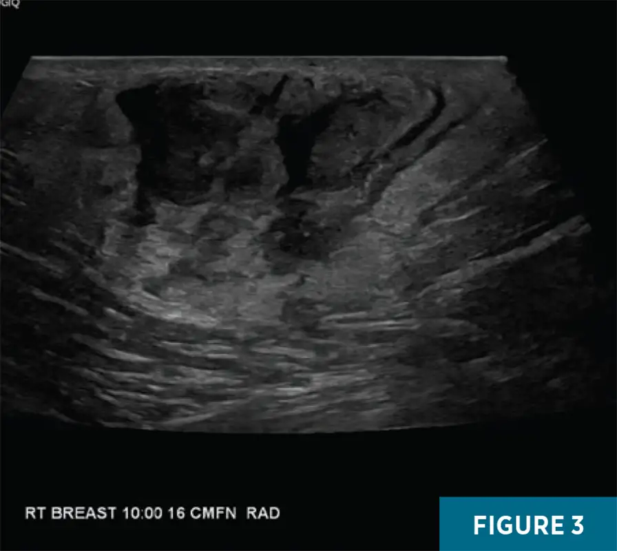
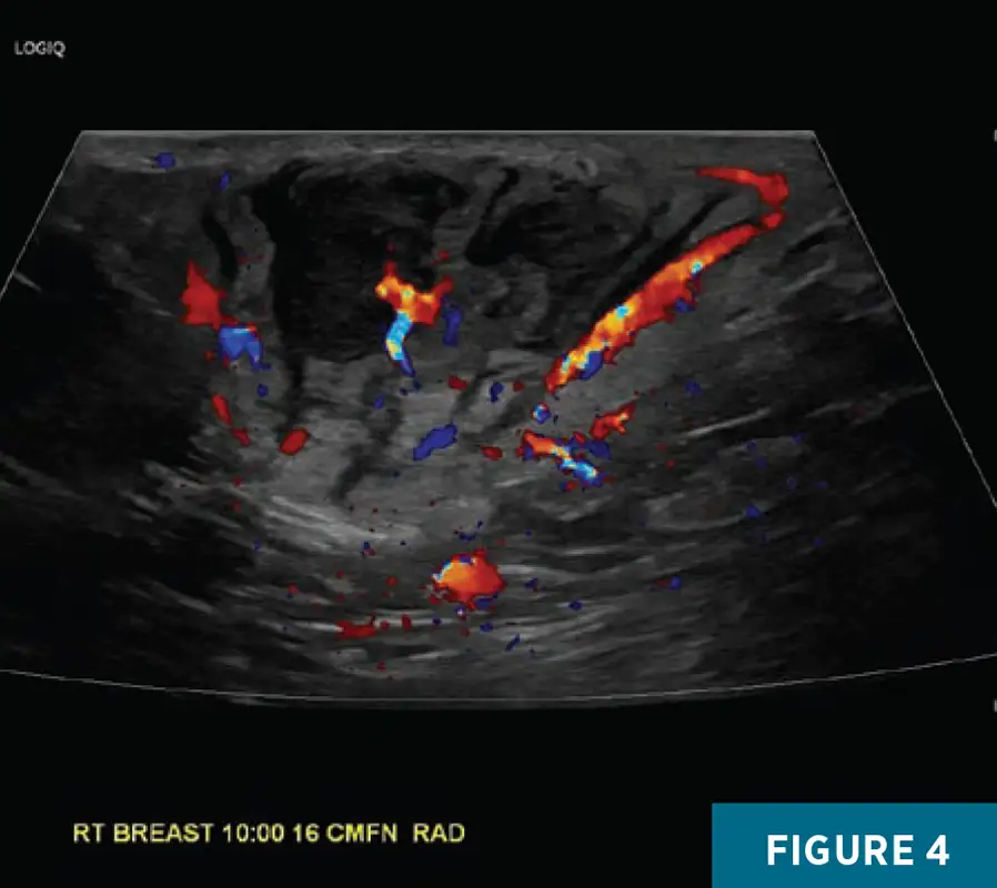
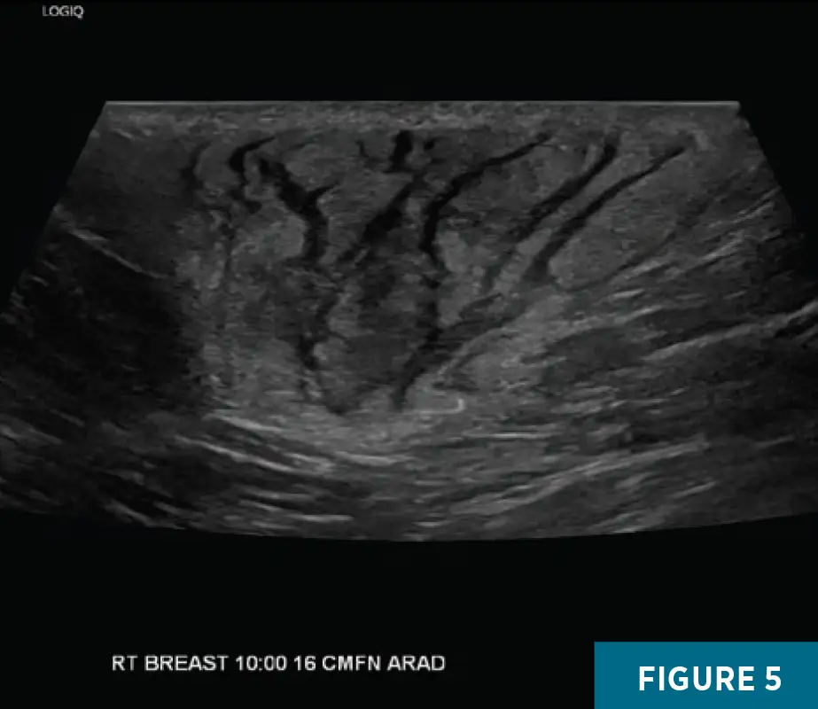
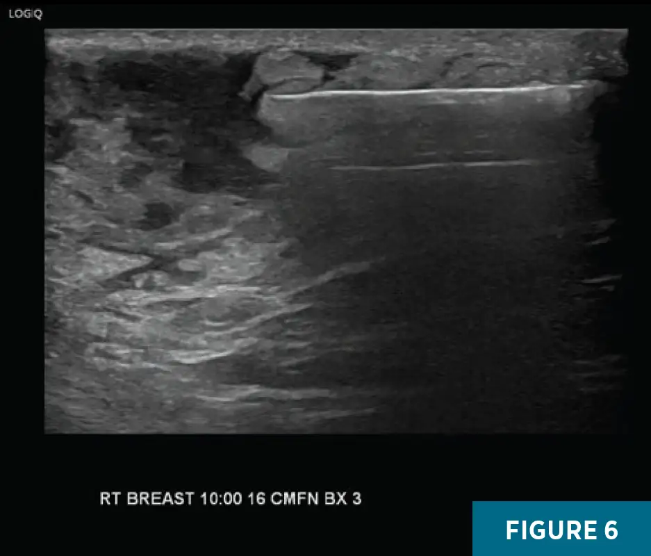
Figures 1, 2, and 3: Diagnostic right breast mammography and targeted ultrasound demonstrated a 5.7 X 4.9 cm focal asymmetry in the posterior third of the right breast, with a corresponding indistinct heterogeneous subcutaneous mass which corelated as palpable and was associated with dermal thickening. Figures 4 and 5: A targeted grayscale ultrasound of the right breast at the 10 o’clock position, 16 cm from the nipple, demonstrated a 5 X 2.4 cm heterogeneous mass involving all depths of the breast and extending from the deep dermis to the chest wall with associated vascularity. Figure 6: An ultrasound image obtained during core biopsy demonstrates the needle within the solid component of the mass.
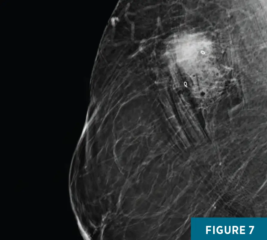
An initial ultrasound-guided core biopsy yielded profound mixed acute and chronic inflammation with no evidence of carcinoma. Immunohistochemistry demonstrated a mixed B- and T-cell infiltrate with scattered histiocytes and plasma cells, negative CD30 and cyclin D1, focal nonspecific S100 staining, and a proliferation index of 5%. Epstein- Barr virus in situ hybridization was negative. These findings did not account for the imaging appearance. Following multidisciplinary review, sampling was considered unrepresentative and repeat ultrasound-guided biopsy of additional solid components was recommended to establish the diagnosis. Figure 7: Postrebiopsy procedure mammography demonstrated two biopsy marker clips in the targeted mass.
Diagnosis
Extranodal Rosai-Dorfman-Destombes disease (RDD) of the breast
Discussion
RDD rarely involves the breast, with roughly 90 cases reported worldwide through 2021.1 A 5 cm mass with indistinct margins spanning dermis to chest wall is a suspicious presentation, and the differential is dominated by malignancy: invasive carcinoma including the inflammatory phenotype, lymphoma, and angiosarcoma. Nonmalignant considerations include abscess or phlegmon, idiopathic granulomatous mastitis, IgG4-related disease, and sarcoidosis; imaging cannot separate these entities, and tissue sampling is required.2
RDD is a non-Langerhans cell histiocytosis, long regarded as reactive and inflammatory. Recurrent somatic MAPK-pathway mutations, most often involving KRAS and MAP2K1 and present in up to one-third of cases, established at least a subset as clonal, and RDD is now classified among histiocytic neoplasms in the fifth edition of the World Health Organization classification of hematolymphoid tumors.3,4 The classic manifestation is massive painless cervical lymphadenopathy; roughly one-quarter of patients have exclusively extranodal disease.2
Mammography in mammary RDD typically shows a high-density mass with indistinct or partially circumscribed margins and no calcifications. Sonographic appearances are variable, reported as hypoechoic, heterogeneous, or mixed, frequently hypervascular, and often involving the dermis or subcutaneous tissue. 1,2 Initial assessments are usually BIRADS category 4 or 5. The present lesion was centrally heterogeneous with marked peripheral echogenicity, a nonclassic appearance underscoring the absence of any specific sonographic pattern.
The diagnostic hinge in this case was radiologic-pathologic discordance. The initial core biopsy returned profound mixed inflammation without carcinoma, interpreted with a panel directed at lymphoproliferative disease. RDD histiocytes characteristically express S100, OCT2, CD68, and CD163; are CD1a-negative; and display emperipolesis, the intact engulfment of lymphocytes within histiocyte cytoplasm.3,5 When the panel is not directed at histiocytes and the sampled tissue is dominated by background inflammation, the diagnosis is readily missed. Because the result did not account for a 5 cm mass extending to the chest wall, repeat targeted sampling established the diagnosis.
Consensus recommendations advise whole-body FDG PET/CT after diagnosis to define extent and assess treatment response.5 Asymptomatic patients may be observed, resection is reasonable for unifocal disease, and patients with KRAS or MEK alterations may respond to cobimetinib.5,6 Prognosis is generally favorable, although outcomes are worse with lower respiratory tract, renal, or hepatic involvement and with coexisting immune disease.5
The teaching point is not that RDD can be suggested prospectively because it cannot. It is that a benign inflammatory result does not close the case on a suspicious breast mass. Concordance assessment belongs to the radiologist, and genuine discordance mandates repeat sampling.
Nadav Mortman, MD, is a radiology resident at UConn Health in Farmington, Connecticut.
Alex Merkulov, MD, is an associate professor of radiology and section head of women’s imaging at UConn Health.
References
1. Iancu G, Gica N, Mustata LM, Panaitescu AM, Vasile D, Peltecu G. Rosai–Dorfman disease: breast involvement—case report and literature review. Medicina (Kaunas). 2021;57(11):1167.
2. Battle B, McIntire P, Babagbemi K, Mema E. Extranodal multifocal Rosai-Dorfman disease of the breast: a case report. Clin Imaging. 2021;71:49-51.
3. Shah AS, Shaikh MJS, Aswani N, et al. Rosai-Dorfman disease: imaging and updates. Radiographics. 2026;46(7):e250179.
4. Khoury JD, Solary E, Abla O, et al. The 5th edition of the World Health Organization classification of haematolymphoid tumours: myeloid and histiocytic/dendritic neoplasms. Leukemia. 2022;36(7):1703-1719.
5. Abla O, Jacobsen E, Picarsic J, et al. Consensus recommendations for the diagnosis and clinical management of Rosai-Dorfman-Destombes disease. Blood. 2018;131(26):2877-2890.
6. Abeykoon JP, Rech KL, Young JR, et al. Outcomes after treatment with cobimetinib in patients with Rosai-Dorfman disease based on KRAS and MEK alteration status. JAMA Oncol. 2022;8(12):1816-1820.