Close Menu
  • Magazine
    • Current Issue
    • Issue Archive
    • Subscribe
  • Topics
    • AI/Machine Learning
    • CT
    • Fluoroscopy/C-Arm
    • General Radiology
    • Interventional Radiology
    • MRI
    • Nuclear Medicine/Molecular Imaging
    • PACS/RIS/Informatics
    • Radiation Oncology
    • Radiology Management
    • Reimbursement & Coding
    • Research News
    • Ultrasound
    • Women’s Imaging
  • E-Newsletter
  • Education
    • ARMRIT Annual Meeting
    • MRI Books
    • Webinars
  • Careers
  • Events
  • Resources
    • Product Directories
    • Resource Listing
    • Reprints
    • Writers’ Guidelines

Join Our Email List

Facebook X (Twitter) LinkedIn
Trending
  • Lending a Hand
  • Whole-Body Makeover
  • Next Phase
  • Beyond Anatomy
  • Editor’s Note: Steps Forward
  • Radiation Safety: Safety Check
  • AI Insights: Balancing the Load
  • Imaging Informatics: Connecting Silos
Thursday, June 18
  • About
  • Contact
  • Advertise
  • Gift Shop
Facebook X (Twitter) LinkedIn
Radiology Today MagazineRadiology Today Magazine
Subscribe
  • Magazine
    • Current Issue
    • Issue Archive
    • Subscribe
  • Topics
    • AI/Machine Learning
    • CT
    • Fluoroscopy/C-Arm
    • General Radiology
    • Interventional Radiology
    • MRI
    • Nuclear Medicine/Molecular Imaging
    • PACS/RIS/Informatics
    • Radiation Oncology
    • Radiology Management
    • Reimbursement & Coding
    • Research News
    • Ultrasound
    • Women’s Imaging
  • E-Newsletter
  • Education
    • ARMRIT Annual Meeting
    • MRI Books
    • Webinars
  • Careers
  • Events
  • Resources
    • Product Directories
    • Resource Listing
    • Reprints
    • Writers’ Guidelines
Radiology Today MagazineRadiology Today Magazine
Home»Issues»April 2014»One the Case

One the Case

Facebook Twitter LinkedIn Email Threads Bluesky Copy Link

By Pedram Parva, MD

History

A 39-year-old woman presented with left third nerve palsy.

Findings

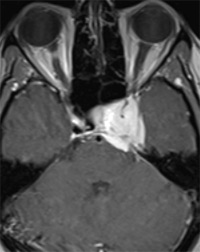
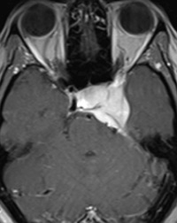
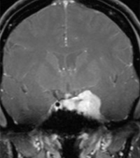
Axial and coronal T1-weighted images after contrast injection showed an intensely enhancing lesion in the left cavernous sinus encasing the intracavernous portion of the left internal carotid artery (compared with the right normal side). This lesion causes cerebellar tentorial thickening (Figures A and B).

A small ipsilateral dural tail can be seen (Figures B and C). The lesion extends to the right cavernous sinus as well as to the left superior orbital fissure (Figure B).

Diagnosis

Cavernous sinus meningioma

Discussion

Meningiomas are the most common primary nonglial intracranial tumors. They mostly affect people in middle age and older, with a 2:1 female dominance.

Typical meningiomas are homogeneous, broad based, and markedly enhancing extra-axial lesions that are firmly attached to the adjacent dura mater. Approximately 50% of meningiomas are parasagittal or attached to the sagittal sinus. Unusual imaging features such as cysts, ring enhancement, or fatty transformation occasionally are seen. The distinguishing characteristic of cavernous sinus meningioma is its predilection to encase the carotid artery, causing stenosis.

Differential Diagnosis

Peripheral nerve sheath tumor, hemangioma, perineural spread, lymphoma, Tolosa Hunt syndrome

Treatment

The main treatment of meningioma is surgical resection. Radiation therapy may be used in cases when surgery isn’t feasible.

— Pedram Parva, MD, is an attending diagnostic radiologist at the Boston University School of Medicine and the VA Healthcare System in Boston as well as an instructor at Harvard Medical School.

Figure AFigure B
 
Figure C 

Resources

  1. Shaman MA, Zak IT, Kupsky WJ. Best cases from the AFIP: involuted sclerotic meningioma. RadioGraphics. 2003;23(3):785-789.
  2. Buetow MP, Buetow PC, Smirniotopoulos JG. Typical, atypical, and misleading features in meningioma. RadioGraphics.1991;11(6):1087-1106.
  3. Bag AK, Shah R. AJR teaching file: cavernous sinus mass in a woman presenting with painful ophthalmoplegia. AJR Am J Roentgenol. 2010;195(3 Suppl):WS1-WS4.

Department
Share. Facebook Twitter Pinterest LinkedIn Tumblr Email

Related Posts

Editor’s Note: Steps Forward

June 1, 2026

Radiation Safety: Safety Check

June 1, 2026

AI Insights: Balancing the Load

June 1, 2026
  • Facebook
  • X
  • LinkedIn

E-Newsletters

A trusted resource for industry professionals, Radiology Today reports the latest news and information that matters to radiologists, radiology administrators, and technologists.

1721 Valley Forge Road #486, Valley Forge, PA 19481
Phone: 800-278-4400 or 610-948-9500
Subscriptions: 833-790-6897

Facebook X (Twitter) LinkedIn

Subscribe

  • Home
  • Subscribe
  • About
  • Contact
  • Advertise
  • Privacy Policy
  • Terms & Conditions
© 2026 Radiology Today Magazine. All rights reserved.

Type above and press Enter to search. Press Esc to cancel.